The brain is split into two halfs called hemispheres. Each of the hemispheres consists of five major parts, the four lobes (frontal, temporal, parietal, and occipital lobe) as well as the cerebellum. They are connected via the brainstem which is another big part of the brain. These components of the brain are described in detail on the following page.

The Lobes
Frontal Lobe
The frontal lobe is the biggest part of the brain. It controls many important abilities including planning and movement as well as social and emotional processes [2] or concentration [1]. The frontal lobe is also responsible for storing and retrieving memories. For all abilities the frontal lobe splits into three major parts, the prefrontal cortex which surrounds Broca's area at the bottom and the dorsolateral prefrontal cortex at the top, the premotor cortex and the primary motor cortex (from front to back).
The prefrontal cortex plays a big role in who we are and how we behave as it reasons on recent memories and acts as the emotional moral compass [1].
Broca's area is part of the language processing center. It strongly correlates with Wernicke's area (see Temporal Lobe). The language is only processed in one half of the brain (mostly the left one even if your left handed). For language processing the Broca's area plans tongue and lip movement [1].
The premotor cortex is responsible for planning the movement of the body. It also saves reaction so if you see a green traffic light for the second time you react faster than the first time [1].
The primary motor cortex acts as motor for the premotor cortex. It performs the planned body movements. The specialty about this is that the right half controls the left body half and the other way round.
Parietal Lobe
The parietal lobe recognizes touch, pressure, temperature, and pain [1]. The pain can be located very precisely because of the lobe. The two halfs have different tasks. The dominant hemisphere is responsible for speech, reading, writing and calculating while the other uses "body memory" to react to the surring environment without having to think about it [1]. The part where the parietal and temporal lobe are connected somewhere arround Wernicke's area is called the temporo-parietal junction and helps to be empathetic. This part also is involved in out-of-body experiences [1].
The front part of the parietal lobe is called somatosensory cortex. It helps humans especially children to learn from objects they touched like a hot stove [1].
Temporal Lobe
The temporal lobe processes the data gathered by the senses (seeing, hearing, feeling, and smelling). It also stores word to sound information [1]. This information is used by Wernicke's area which strongly works togehter with Broca's area (see Frontal Lobe).
The lobe is also included in the visual memory process for recognizing objects and persons like our favorite place or our best friend's face [2].
Occipital Lobe
The occipital lobe is the smallest of the four lobes. It uses data from the eyes to determine shape, color, depth, and angles [1]. For visual processing it works together with other parts of the brain like the Temporal Lobe.
The inner part of the lobe is called visual cortex. It uses gathered information and spreads them to the corresponding area in other lobes of the brain to set objects in relation to your body or calculate its movement speed and direction [1].
The Cerebellum
The cerebellum, also known as "little brain", is important for balancing and learning. It enables us to ride a bike [2] or to hit a baseball [1]. It takes around 10% of the brain's total weight and contains more neurons than the rest of the brain together [3]. The outer layer contains gray matter while the inside is full of white matter [1]. The gray matter is a cluster of neuron dendrites while white matter is the bundle of axons the so called tail of a neuron (see also neurons) [6]. In the center of the cerebellum there is a small nuclei which is connected to the motor cortex in the frontal lobe [1].
The Brainstem
The brainstem is responsible for autimatics and reflexes like breathing, blood pressure and heart rate [3]. It consists of four "floors".
The topmost level is the Diencephalon which is between the two hemispheres of the brain at the height of the eyes. It contains the third ventricle, the thalamus and hypothalamus [1].
The next floor from the top is the mid-brain. Nerve fibres are extended upwards to the cerebrum (the lobes) and downwards to the spinal cord [1].
On their way down they pass the pons which controls the sleep-wake rhythm [1].
All bundeled nerves go through the medulla oblangata, the lowest level of the brainstem, to the spinal cord [1].
Ventricle
A ventricle is a open space within the brain channeling the cerebrospinal fluid (CSF) [1]. There are four ventricles, two lateral, the third and the fourth ventricle.

The lateral ventricles lead to the two hemispheres. The third ventricle is bounded by the thalamus and the hypothalamus [1]. The fourth ventricle is connected via the cerebral aqueduct as shown in the image. It is the lowest ventricle and lies between the midbrain, pons and medulla oblangata.
Thalamus
The thalamus is a big distributor. It receives plenty of information gathered by the senses. Afterwards, it decides what information is relevant and sends it to the cerebrum [1]. A similar action is done with emotional signals received from the hypothalamus, but these are sent to the limbic system. Thereby, the sleep of a human is influenced [1].
Hypothalamus
The hypothalamus is located in the midbrain between the two hemispheres. It is just as big as the end of your little finger. It controls autonomic nervous systems which includes blood pressure, pulse, sweating and more [1]. It also controls the endocrine systems which regulates groth, metabolism and reproduction with the help of hormones [1]. The hypothalamus tries to maintain a stable balance called homeostasis.
The 12 Crainial Nerves
The 12 crainial nerves make us aware of our environment and enable the interaction [1]. They serve the specified body parts. The table content was extracted from [1].
| I. | Olfactory | smell |
| II. | Optic | sight |
| III. | Oculomotor | eye (up and down) and eyelid movement, pupils control |
| IV. | Trochlear | move eyes towards nose |
| V. | Trigeminal | chewing (jaw movement), sensation in eyes, teeth and face |
| VI. | Abducens | move eyes away from nose |
| VII. | Facial | tear and salvia production, taste (tip and middle of tongue), facial expressions |
| VIII. | Vestibulocochlear | hearing and balance |
| IX. | Glossopharyngeal | taste (back of tongue), touch, temperature in mouth, tongue and throat movement,swallowing, gag reflex |
| X. | Vargus | swallowing, breathing, heart rate, stomach acid production |
| XI. | Spinal accessory | move head, neck and shoulders, larynx and throat (swallowing) |
| XII. | Hypoglossal | move tongue |
Neurons
The brain consists of blocks of neurons. They transport information through the body using electrical and chemical signals [4]. A neuron is constructed as follows.

A neuron is split into two major parts, a receiving and calculation part and a conducting and transmitting part [4]. The information transmitted depends on the location in the nervous system.
Dendrites (left purple part in the image) create synapses. With these, a connection to the target cell is established. The axon (yellow part in the image) is the so called tail of a neuron and conducts and transmits information. In some special cases the axon receives information [4].
Protection of the Brain
The Meninges
The meninges covers the brain from damage and is right under the cranium (bone). In the image the three layers, the dura mater, the arachnoid mater and the pia mater, can be seen.

The dura mater is the outermost layer consisting of two connective layers. The endosteel layer connects the dura mater with the bone of the skull [5]. The second connective layer is the meningeal layer. It connects the the endosteel layer with the middle layer of the meninges. Underneath the arachnoid is the sub-arachnoid space. It contains cerebrospinal fluid [5]. The pia mater is the innermost layer. It is very thin and really near to the surface of the brain [5].
Cerebrospinal Fluid (CSF)
The cerebrospinal fluid is a clear, colorless liquid that surrounds the brain. At a time there are between 100 and 160 ml CSF [6]. Per day arround 500 ml are absorbed by the brain [6] and are produced within the walls of the ventricles [1] which distribute the CSF all around the brain. Its functions are buoyancy so the pressure does not damage the nervous system and the brain is not aggrieved by its own weight [6]. Another function is the protection from shaking the head so the brain does not touch the cranium. If it is shaken too hard it leads to a concussion or at children to the shaken child syndrome [6].
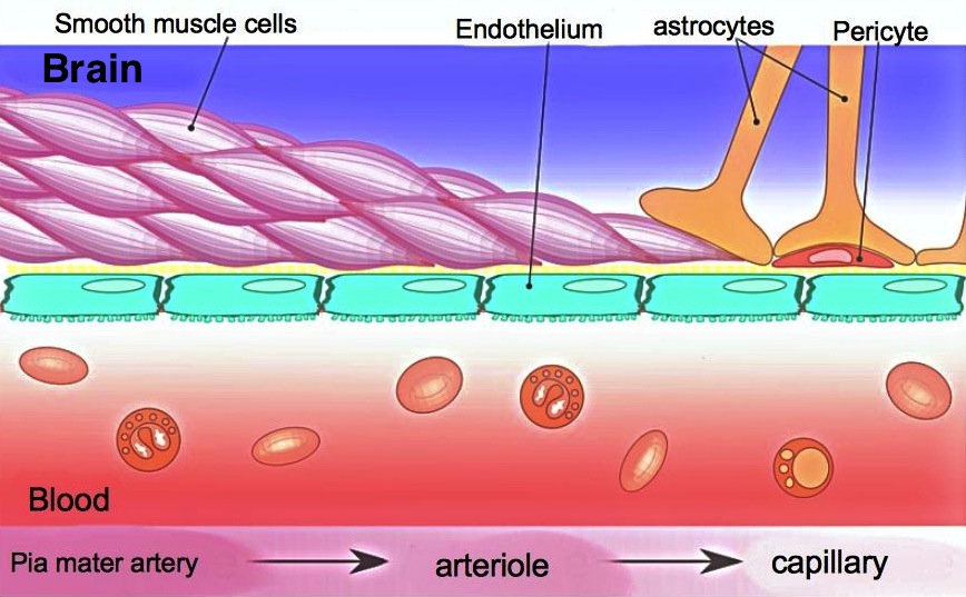
Blood-Brain Barrier
The blood-brain barrier is a semipermeable membrane barrier which distinguishes what substances can enter the brain. Antibodies for examples are too large to pass the endothelium cells and also only certain antibiotics can pass which makes the treatment of infections very difficult [7].
Bibliography
- http://www.finr.net/files/brain/index.htm
- https://www.koshland-science-museum.org/explore-the-science/life-lab/brain#anatomy
- http://www.g2conline.org/2022
- https://owlcation.com/stem/Structure-of-a-Neuron
- http://teachmeanatomy.info/neuro/structures/meninges/
- https://www.slideshare.net/nicolezemroz/chapt14-lecture-4
- https://en.wikipedia.org/wiki/Blood–brain_barrier
- https://upload.wikimedia.org/wikipedia/commons/1/12/Blood_vessels_brain_english.jpg
Kommentar
Unbekannter Benutzer (ga47xoj) sagt:
16. Mai 2017Feedback from Group 3:
Rest of the page looks good. Good work