This subpart focuses on different brain tumor types. After looking at classification based on cell behaviour and cell origin, we look at some of the most common primary brain tumors. In the end we give a brief overview of the WHO classification of brain tumors.
Classification
On a basic level tumors are classified based on their cell origin and their behaviour. However tumors can have different subtypes which show different behaviour, which leads to a great variety of idividual brain tumors.
Based on cell behaviour:
Benign brain tumors contain cells which grow differently than normal tissue cells but which are not cancerous. These tumors grow slowly, rarely spread to the neighbouring tissue and have a well-defined border. Mostly they can be removed by surgery and will not reoccur afterwards. However they can become dangerous when they press on certain parts of the brain and compress normal brain tissue.
Malignant brain tumors contain cancerous cells which grow rapidly compared to normal brain cells. They can spread to healthy brain tissue nearby invading and destroying it. These tumors have a diffuse border. They mostly don't spread outside the brain. On the other side many malignant tumors are secondary tumors originating from other parts of the body.
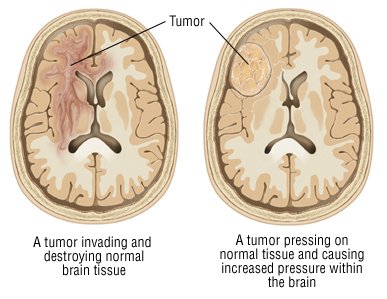
Figure 1: Illustration of a begnin tumor (right) and a malignant tumor (left) [10]
Based on cell origin:
Primary tumors originate from the brain tissue and stay there. They can be benign or malignant. In contrast, secondary tumors arise in other parts of the body (e.g. the lung or the skin) and spread into the brain. This type is mostly malignant and more common than primary tumors.
Primary brain tumors
According to the American Brain Tumor Association from January 2017, the most common primary brain tumors arise as follows [5]:
- Meningioma: 36,6%
- Gliomas: 24,7% (74,6% of all malignant tumors)
- Glioblastomas: 14,9% (55,4% of all gliomas)
- Astrocytomas represent 75% of all gliomas (glioblastoma included)
- Pituitary tumors: 16%
- Nerve sheat tumors: 8,2%
- Lymphomas: 2%
- Oligodendrogliomas: 2%
- inside the brain primary tumors are most common in the meninges (37%)
Meningiomas
Meningiomas arise from the meninges. These are three tissue layers that cover the brain and the spinal cord. So the tumors are mostly located at the border of the brain. Tumors of this type are mostly benign and slow growing. So they don't need immediate treatment and can often be removed by surgery. However they still can become dangerous if they grow too big and compress healthy brain tissue by expanding. [6]

Figure 2: locations of meninigiomas [6]
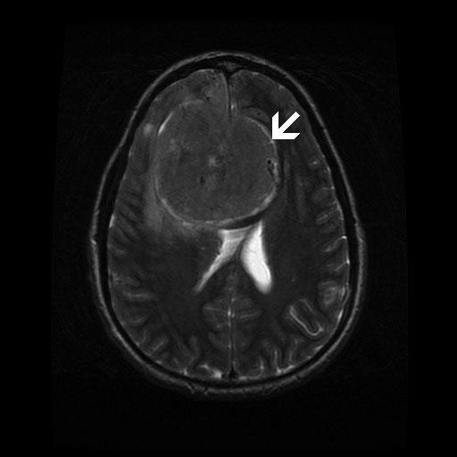
Figure 3: T2-weighted image with large meningioma in the top center [11]
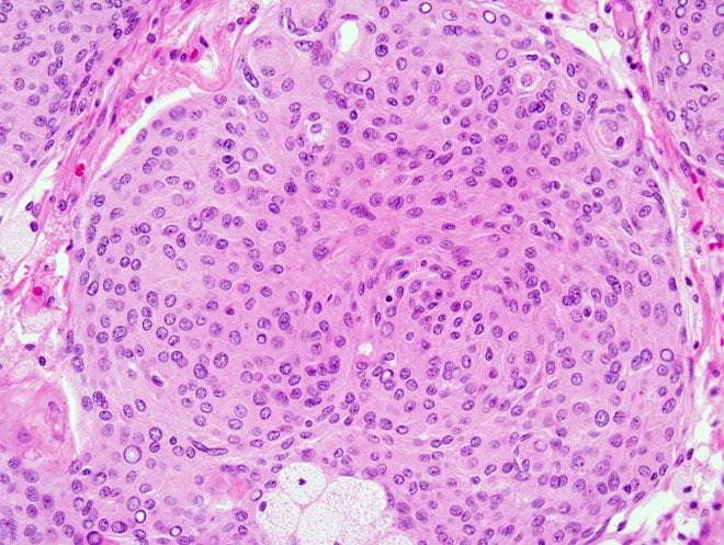
Figure 4: microscopic view of a grade I meningothelial meningioma [11]
Glioblastoma
Glioblastomas, or Glioblastoma multiforme (GBM), arise from astrocytes, which are a form of glial cells. The former ones are star-shaped cells that support the neurons. Due to its high reproducing rate and support of blood vessels, this type of tumor is highly malignant. The fast growth also leads to a decrease of oxygen supply in the tumor center and therefore cells in the center of the tumor die (necrosis). The invasive behaviour of this tumor also leads to very diffusive borders. [7]
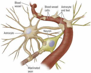
Figure 5: schematic figure of an astrocyte [12]
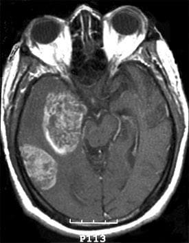
Figure 6: T1-wegihted axial MRI showing a multicentric GBM [13]
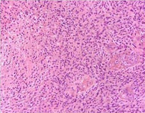
Figure 7: histopathological slide showing GBM [13]
Astrocytomas
Astrocytomas develope from astrocytes as well. They can be classified based on the grading system explained in Group 6 - Tumor Grading and show group-specific behaviour. Examples for the different groups include:
- Grade I: Pilocytic Astrocytoma; these tumors grow slowly and typically don't spread, however they can still become noticably big.
- Grade II: Diffuse Astrocytoma; these tumors grow slowly but also invade surrounding healthy brain tissue, subgroups of this type are formed regarding appearance and behaviour of the tumor cells.
- Grade III: Anaplastic Astrocytoma; these tumors can have outer parts which look like strings invading the surrounding tissue.
- Grade IV: Glioblastoma
Oligodendroglioma
These tumors are a type of glioma and origin from oligodendrocytes which are glial cells that support and insulate axons [11]. Tumors of this type can be classified as grade II or anaplastic grade III and are located in the cerebral hemisphere (mostly the frontal and the temporal lobes). While grade II ones rather show a round and compact form, grade III oligodendrogliomas also show vascular proliferation and can therefore exhibit a more deformed shape. [9]

Figure 8: schematic figure of an oligodendrocyte [14]
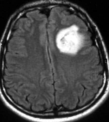
Figure 9: axial MRI of a low-grade oligodendroglioma [15]

Figure 10: mircographic view with red blood vessels, cells in dark purple and well-defined cell borders [16]
WHO classification
The following table shows the 2016 WHO classification of tumors of the central nervous system.


Bibliography
[1] http://www.webmd.com/cancer/brain-cancer/brain-tumor-types#1
[2] http://www.healthline.com/health/brain-tumor#overview1
[3] http://braintumor.org/brain-tumor-information/understanding-brain-tumors/tumor-types/#Meningioma
[4] http://www.medicinenet.com/brain_tumor_symptoms/views.htm
[5] http://www.abta.org/about-us/news/brain-tumor-statistics/
[6] http://www.abta.org/brain-tumor-information/types-of-tumors/meningioma.html
[7] http://www.abta.org/brain-tumor-information/types-of-tumors/glioblastoma.html
[8] http://www.abta.org/brain-tumor-information/types-of-tumors/astrocytoma.html
[9] http://www.abta.org/brain-tumor-information/types-of-tumors/oligodendroglioma.html
[10] https://www.drugs.com/health-guide/brain-tumor.html
[11] http://emedicine.medscape.com/article/1744164-overview#a5
[12] http://jonlieffmd.com/blog/astrocytes-control-synapse-function
[13] http://emedicine.medscape.com/article/283252-overview
[14] http://medical-dictionary.thefreedictionary.com/oligodendrocyte
[15] http://emedicine.medscape.com/article/342958-overview#a1
2 Kommentare
Unbekannter Benutzer (ga46zuy) sagt:
16. Mai 2017The page is very informative. You could add a differentation between tumor and cancer, and a bibliography.
Otherwise good job !
I like that you used a lot of pictures
Unbekannter Benutzer (ga67yur) sagt:
16. Mai 2017Thanks for the feedback! We will work on a bibliography.