This part deals with the grading scheme for brain tumors.
According to the World Health Organization (WHO), brain tumors can be graded depending on histological features (showing the microanatomy of cells and tissue [1]) and the tumors malignancy. This results in four different groups. Grade I and II are also called low-grade tumors, while Grade III and IV are high-grade tumors. Over time low-grade tumors can evolve to high-grade ones. The following list shows characteristic features of each group [2] [3].
- Grade I: These tumors are least malignant and slow growing. The cells appear nearly normal. Surgery can be an effective treatment and these tumors are often associated with long-term survival.
- Grade II: Tumors of this group are slightly more malignant. They still grow relatively slowly, but the cells look abnormal to some extent and the cells can spread to and recur in nearby tissue.
- Grade III: These tumors are malignant. They reproduce abnormal cells and grow into nearby normal brain tissue. Tumors of this grade often recur as Grade IV tumors.
- Grade IV: These are the most malignant tumors. Their cells reproduce rapidly and can grow into nearby normal tissue fast. They are able to form new blood vessels (angiogenesis) to maintain the rapid growth. However a loss of oxygen also leads to dead tumor cells in the center of these tumors (necrosis). The most common tumor type in this group is glioblastoma multiforme.
To determine the grade of the tumor, samples from a biopsy are analysed. If these samples contain cells of different groups, the highest grade of the tumor cells also defines the grade of the tumor. Features of grade IV tumors, such as necrotic cells, can also be seen on medical images. So this is another way to estimate the grade of a tumor on first sight.

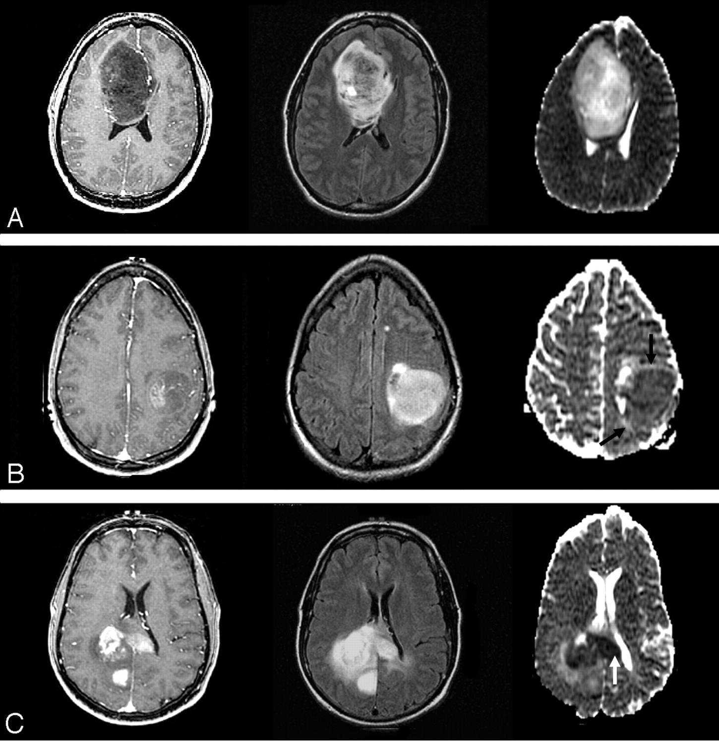
Figure 1: T1- weighted (left), FLAIR (middle) images and ADC maps (right) of grade II (A), grade III (B) and grade IV (C) astrocytomas [5]
2 Kommentare
Unbekannter Benutzer (ga46zuy) sagt:
16. Mai 2017Compact layout with all the necessary information. It's a bit misleading the the image caption one time is above and the other time is beneath the image.
Also your first link is a 3, maybe change it to the right order. You could also add a bibliography at the end of the page
Nonetheless, good work
Unbekannter Benutzer (ga67yur) sagt:
16. Mai 2017Thanks for the feedback. You're right, I will adjust the caption and the links, thanks for noticing And we will work on the bibliography.