Motivation
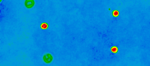
For the simplification and acceleration of blood analyses, flow cytometry microscope was crafted for the researchers at TranslaTUM. Digital holography allows us to take 3D images of any kind of cells pumped through the microfluidic channel. Since the different components of the Cell, for example cell wall or cell nucleus, different refractive indices the light needs different long to cross this one. This phase difference is then shown as an interference pattern visible in the hologram. In order to make the resulting images interpretable the goal of the project proposed here is a software prototype, which consists of two closely is made up of the main components connected with each other.
Road Map
Data Warehouse
Lorem ipsum dolor sit amet, consetetur sadipscing elitr, sed diam nonumy eirmod tempor invidunt ut labore et dolore magna aliquyam erat, sed diam voluptua. At vero eos et accusam et justo duo dolores et ea rebum. Stet clita kasd gubergren, no sea takimata sanctus est Lorem ipsum dolor sit amet. Lorem ipsum dolor sit amet, consetetur sadipscing elitr, sed diam nonumy eirmod tempor invidunt ut labore et dolore magna aliquyam erat, sed diam voluptua. At vero eos et accusam et justo duo dolores et ea rebum. Stet clita kasd gubergren, no sea takimata sanctus est Lorem ipsum dolor sit amet.
Algorithms
This stage is a Machine Learning Toolbox, which is based on the current research results in the area of Deep Learning for image processing, object recognition, classification and image segmentation. Classical methods from image processing, dimension reduction and feature extraction complete the selection of the required tools. The objective for this component is the robust and efficient acquisition of information about the digitized cells. Some research groups are already pursuing this approach, but a machine learning tool is only useful if it is trustworthy and accepted. Clinical relevance can only be achieved if the tool is intuitive to use and provides clear statements. In addition, it must be characterized by statistical significance, robustness and reliability, which can only be achieved through frequent successful use. Therefore, the direct involvement of colleagues from the fields of medicine and biology plays a decisive role. The users of the software are actively involved from the project conception phase onwards, as this is the only way to ensure successful integration into clinical operations.
Collaboration Platform
This component is based on the requirements and knowledge of the users. It consists of an intuitive user interface that makes the procedures from the toolbox transparent and coordinates their use. In addition, database accesses are controlled and the compilation of the required data sets is facilitated. To increase the significance of the results from the toolbox, the user interface supports the presentation of large amounts of data and possibilities for statistical analysis. As is well known from machine learning research, the quantity and quality of the available data is a decisive factor for the success of the methods used. Therefore, the software prototype offers the possibility to quickly generate a sufficient amount of labelled data through Human Assisted Labeling (the AI makes suggestions, the human validates). A suitable goal for this component is thus a practical implementation of medical and biological expertise in the algorithms and successful use as a clinical assistance system.
Clinical Decission Support System
Lorem ipsum dolor sit amet, consetetur sadipscing elitr, sed diam nonumy eirmod tempor invidunt ut labore et dolore magna aliquyam erat, sed diam voluptua. At vero eos et accusam et justo duo dolores et ea rebum. Stet clita kasd gubergren, no sea takimata sanctus est Lorem ipsum dolor sit amet. Lorem ipsum dolor sit amet, consetetur sadipscing elitr, sed diam nonumy eirmod tempor invidunt ut labore et dolore magna aliquyam erat, sed diam voluptua. At vero eos et accusam et justo duo dolores et ea rebum. Stet clita kasd gubergren, no sea takimata sanctus est Lorem ipsum dolor sit amet. Lorem ipsum dolor sit amet, consetetur sadipscing elitr, sed diam nonumy eirmod tempor invidunt ut labore et dolore magna aliquyam erat, sed diam voluptua. At vero eos et accusam et justo duo dolores et ea rebum. Stet clita kasd gubergren, no sea takimata sanctus est Lorem ipsum dolor sit amet.
Translation Approach
Approximately 480,000 people per year fall ill with cancer in Germany. Cancer is also the second most frequent cause of death. The improvement of diagnostic and therapeutic options is therefore an important task of cancer research. At TranslaTUM, scientists from medicine, engineering and the natural sciences work together to improve the chances of curing cancer patients. Therefore, the overarching idea of this project is to quickly transfer novel findings from research into clinical practice - a process known as translation.Different diseases leave traces on cells, in particular on blood cells, in different ways. Therefore, modern haematology analysers should be able to detect a variety of abnormalities and changes in the morphological structure and composition of cells and blood. Since the irregularities observed can sometimes be very rare, the cells must be examined in high throughput to obtain a statistically significant result. Such a system is also expected to be self-sufficient, especially for use in remote areas where the so-called neglected or poverty-prone diseases are still common. The duration of the analysis should be short, not only in urgent cases, so that the process can be kept as simple as possible for the doctor and the patient. Even if a system can provide the services mentioned, it must still meet at least three more criteria in order to achieve clinical relevance. First and foremost, it must meet high safety and quality standards. In addition, the approaches used must be explainable, as black box solutions are largely unacceptable in the treatment of humans and animals. Last but not least, the user-friendliness of the software and devices developed plays a decisive role in ensuring that they can be used productively. Finally, technological progress is aimed at an end-to-end approach that makes it possible to support physicians in the diagnosis and therapy of a large number of diseases with only a single patient sample.
As science and technology are still far away from such a comprehensive and flexible haematology analyzer, this project will take a step in the indicated direction. As described, DHM in combination with a microfluidic channel allows a high throughput of cells and at the same time a relatively detailed view of the cell's composition and inner workings. Diseases like leukemia can not only destroy the natural balance of white blood cells, but also cause the release of unfinished precursor cells. Therefore, not only the normal subgroups have to be distinguished and counted, but also the special cases, which is not possible with conventional haematology analysers. Another example is pancreatic cancer. In this case, it is important to know at which stage the cancer cells are located in order to be able to develop and evaluate appropriate chemotherapies. Therefore, subtle deviations in the shape of the cells must be detected. In general, this problem also exists in the differentiation and purification of cell cultures, which is hardly possible for the human eye without sample preparation. In general, the image-based detection, classification and analysis of cellular objects offers great potential in the fight against intra- and extracellular parasites. In the case of drug development for schistosomiasis, the aim is to monitor the viability of the worm-like parasites. The same applies to the detection and treatment of malaria, where plasmodia attack red blood cells.


