Brain Anatomy
The Lobes

3 major parts:
- Cerebrum
- Cerebellum
- Brainstem (diencephalon, midbrain, pons, medulla oblongata)
Useful links:
- Lobes along with some related functions [4]
- Interactive brain view [5] and [6]
- Interactive brain view, associated functions, disorders, ... [7] and in an app (search for 3D Brain in any app store)
Fresh brain, anatomy explained. WARNING: Graphic images of human brain
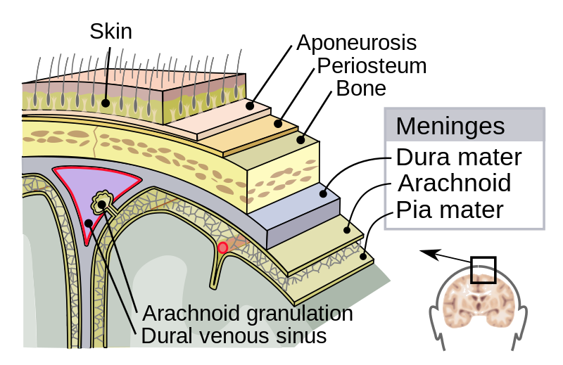
The meninges - protection of the brain


image source: [2] (left), [3] (right)
Meninges are connective tissue membranes enveloping the brain → protect brain, provide structural framework for blood vessels
Opening the dura mater induces brain shift
CSF - more protection [8,9]
Cerebrospinal fluid (CSF) acts as buffer for the brain inside the skull.
 Pulsating CSF shown in MRI [8]
Location
- between arachnoid and pia mater in brain and spine
- in ventricular system (inside and around brain and spinal cord
Function
- Buoyancy
- Protection
- Decrease intracranial pressure
- Homeostasis (keep steady levels of e.g. glycine)
- Clearing waste (e.g. metabolic toxins)
Blood-brain barrier [9]
Steady blood supply to neurons has to be given (need for ATP, glucose, oxygen)
Without blood:
- 10 s: loss of consciousness
- 2 min: impairment of neural function (talking, weekness, paralysis)
- 4 min: irreversible brain damage
Tight endothelial cells of the brain barrier keeps harmful substances (bacteria, antibodies, ...) out and lets important things like ATP, glucose, lipid-soluble substances (O2, CO2, alcohol, nicotine, anesthetics ) pass.
What else?
- Functional areas of the brain → different behaviour to tumors located there
- Giving directions. What is "rostral", "caudal", ...?
- Gray (cell bodies, dendrites and synapses) and white (axons) matter → what does a neuron look like
- Ventricles
Brain Tumor Symptoms [10]
- Headaches
- Seizures
- Sensory (touch) and motor (movement control) loss
- Deep venous thrombosis (blood clot)
- Hearing loss
- Vision loss
- Fatigue
- Depression
- Behavioral and cognitive (thinking) changes
- Endocrine dysfunction (hormone/gland changes)
What else?
- Relate symptoms to functional areas and/or tumor type
- Can certain features of otherwise unspecific symptoms be an indicator? (e.g. headache - may worsen with coughing, exercise, or a change in body position.)
Brain Tumor Diagnosis [10]
Neurological exam
Assess patient's nerves, senses, muscle strength, reflexes, balance, and mental state.
Tests include: vision, hearing, reflexes, balance, sense of touch and smell, abstract thinking ability and memory.
Brain scans
With or without contrast agent possible
Anatomical imaging
Computed Tomography (CT)
Series of x-ray images
 CT of patient with glioblastoma: [11]
Magnetic Resonance Imaging (MRI)
Several very different sequences, like T1,T2, ...


T1 image with mengiome [12] and T2 image [13]
Dynamic CT or Dynamic MRI
Angiography and MRI Angiography (MRA)
Functional imaging
Magnetoencephalography (MEG)
Measures magnetic activity in the brain that is caused by neuronal activity.
Can be overlayed on top of MRI to identify functional areas in the brain.
Positron Emission Tomography (PET, FDG-PET)
Functional MRI (fMRI)
Diffusion weighted imaging (DWI) & Diffusion tensor imaging (DTI)
Single Photon Emission Computerized Tomography (SPECT)
Magnetic Resonance Spectroscopy (MRS) or Nuclear Magnetic Resonance (NMR)
Biochemical information (such as distribution of alanine in the brain)
Flow-Sensitive MRI (FS MRI)
Lab tests
Lumbar Puncture (Spinal Tap)
Collect CSF by inserting a needle into the spinal canal. Look for tumor cells, blood or proteins (infection) in the sample.
Evoked-potentials
Senses are stimulated (visual, acoustic, ...) and neuronal reaction is measured with EEG.

Audiometry
Endocrine Evaluation
Perimetry
Biomarker Research
DNA profiling
Blood test or biopsy / tissue sample (if solid tumor) to test biomarkers and find which genes are up- or down-regulated. Create personalized treatment plan that fits the profile of specific cancer.
Biopsy
What else?
- Why are these scans taken
- What do they show best (soft tissue, increased activity, ...)
Tumor Types [14]
Primary brain tumor
- Start in the brain and usually do not spread to other organs
- They are rather uncommon
Secondary brain tumor
- More common form
- Start in other parts of the body (often lung, breast, kidney, skin) and spread to the brain
Benign tumor
- Only contain non-cancerous cells
- Clear defined border
- Slow growth rate
- Usually do not spread
- Only operated on if symptoms occur
- Still can be deadly
Malignant tumor
- Contain cancerous cells
- varying growth rate
- sometimes spread
What else?
- Astrocytomas
- Meningiomas
- Oligodendrogliomas
- http://braintumor.org/brain-tumor-information/understanding-brain-tumors/tumor-types/
- http://www.abta.org/brain-tumor-information/types-of-tumors/
Tumor Grading [14]
- Grade 1. The cells look nearly normal and grow slowly. Long-term survival is likely. Can often be cured with surgery
- Grade 2. The cells look slightly abnormal and grow slowly. No necrosis or actively dividing cells. The tumor may spread to nearby tissue and can recur later, maybe at a more life-threatening grade.
- Grade 3. The cells look abnormal and are actively growing into nearby brain tissue. These tumors tend to recur.
- Grade 4. The cells look most abnormal and grow and spread quickly. The tumor has blood vessel growth and necrosis.
What else?
- Definition of tumor grades
- How is it determined
Bibliography
1) https://en.wikipedia.org/wiki/File:Blausen_0101_Brain_LateralView.png
2) https://upload.wikimedia.org/wikipedia/commons/thumb/8/8e/Meninges-en.svg/800px-Meninges-en.svg.png
3) https://upload.wikimedia.org/wikipedia/commons/f/f8/Gray769.png
4) https://www.koshland-science-museum.org/explore-the-science/life-lab/brain#anatomy
5) https://www.koshland-science-museum.org/explore-the-science/interactives/brain-anatomy
6) http://www.finr.net/files/brain/index.htm
7) http://www.g2conline.org/2022
8) https://en.wikipedia.org/wiki/Cerebrospinal_fluid
9) https://www.slideshare.net/nicolezemroz/chapt14-lecture-4
11) https://commons.wikimedia.org/wiki/File:TAC_Brain_tumor_glioblastoma-Coronal_plane.gif
12) https://upload.wikimedia.org/wikipedia/commons/c/cb/Falxmeningeom_MRT_T1_mit_Kontrastmittel.jpg
13) https://en.wikipedia.org/wiki/File:Brain-T2-axial.png
14) http://www.webmd.com/cancer/brain-cancer/brain-tumor-types#1
Kommentar
Julia Rackerseder sagt:
26. April 2017You could add more details!!!
But I really like your wiki