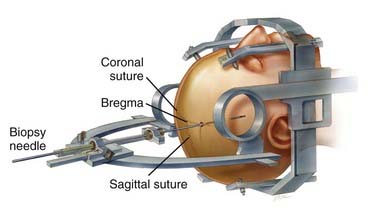
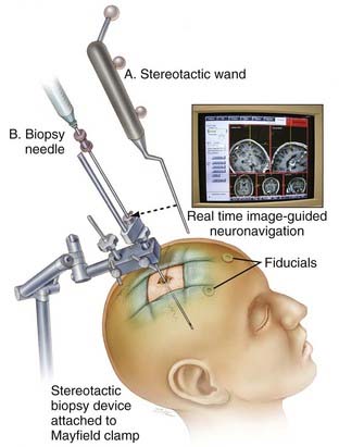
An approach to navigation in neurosurgery relies on frame-based and frameless techniques. Both are used to target structures onto an human head with the 3D-coordinates acquired from preoperative imaging.Therefore the relationship between the coordinate space for the preoperative images and that for the surgical field must be calculated. Frame-Based systems use the same frame for preoperative images and the surgery where the relationship of the two coordinate systems is known. In frameless systems point-pair registration or surface contour registration can be used. This frame principle is also as stereotactic radiation therapy explained in Radiation Therapy. This chapter covers both methods and in the end compares them.
Different systems are used to track the fiducial markers including computer vision and magnetic systems. There is the possibility to use acoustic systems, however the require complex corretion and are susceptible to interference. Optical Systems have the disadavantage that they always require a line of sight. Magnetic systems do not need to see anything but can be disturbed by metallic objects in the operating room. Since optimal placement of this markers can be crucial [6] developed an genetic algorithm to calculate optimal positions of the marker in order to minimize the target registration error. The correspoding marker positions are then shown to the surgeons on the 3D model to help in placement of them.
Comparison
[2] concluded that there is no difference in accuracy using frameless and frame-based markers. However the more recent study [1] suggests that there is a difference, though it is not that big. Both papers recommend to the surgeon to use the method he is most comfortable with.
Comparison of frame-based and frame-less techniques with regard to the diagnostic yield of the biopsy [Source]
There is currently research going on that tries to combine both approaches as suggested in [3]. They propose a head-mounted robot-guided approach that combines the stability of a bone-mounted setup with the flexibility and tolerability of a frameless system. By reducing human interference e.g. manual parameter settings this technology might be particularly useful in neurosurgical interventions that necessitate multiple trajectories.
Bibliography
- Yi Lu et. al. "Comparative Effectiveness of Frame-based, Frameless and Intraoperative MRI Guided Brain Biopsy Techniques" available here
- Dammers R et. al. "Safety and efficacy of frameless and frame-based intracranial biopsy techniques" available here
- F. Grimm et. al. "Blurring the boundaries between frame-based and frameless stereotaxy: feasibility study for brain biopsies performed with the used of a head-mounted robot" available here.
- https://clinicalgate.com/frame-and-frameless-stereotactic-brain-biopsy/#f0020
- http://what-when-how.com/stereotactic-and-functional-neurosurgery/frameless-stereotactic-systems-general-considerations/
- http://medicaldevices.asmedigitalcollection.asme.org/article.aspx?articleid=1451820


