In this part we talk about different common methods that are used to treat brain tumors. Next to describing the methods, we also discuss when they are used and what possible risks might occur. Choosing a method or a combination of different methods depends on patient conditions (e.g. age, health) as well as properties of the tumor that is being treated (e.g location, size, growth, grade). Furthermore the possible benefits and risks of different treatment methods have to be evaluated. To find the right treatment different specialists can be consulted, these include neurosurgeons, oncologists and mental health counselors. [1]
Monitoring
Monitoring via different scanning methods is done for all tumor treatments. However monitoring alone is used for low grade tumors which are not aggressive and for which the patient doesn't show any symptoms. After surgery of these low grade tumors and high grade tumors they are monitored over time to examine whether the tumor recurs. [2]
Surgery
Surgery for brain tumor patients can be performed for different reasons, these include getting samples of the tumor for analysis, removing as much of the tumor as possible, preparation for chemo- or radiotherapy and relieve healthy brain tissue that is affected by pressure from the tumor. Surgery often is the first step in the treatment of brain tumors and is therefore performed for most begnin and many malignant tumors. Medical images (as CT or MRI) are produced before surgery so that the surgeon can plan the procedure efficiently regarding size, location and form of the tumor.

Different types of brain surgery include:
- biopsy: removal of a sample of the tumor that is analyzed later on
- craniotomy: A portion of the skull is removed to cut out as much of the tumor as possible. Afterwards the cut-out portion of the skull is replaced and the skull is closed again.
- craniectomy: Similar to a craniotomy but the part of skull that was removed isn't replaced. This is done when the surgeon expects the brain to bulge after surgery. The bone that was cut out can be stored and replaced later on.
- debulking: removing as much of the tumor as possible
- removal: During partial removal, only parts of the tumor are removed because the removal of other parts would lead to brain damage. The remaining tumor is then treated with other methods. A complete removal, also called "gross total resection", removes all visible parts of the tumor. However some tumor cells can still remain and grow again because they are too small to be seen during surgery.
- shunt: This is a drainage system that is used if too much fluid accumulates in the brain. By inserting a shunt the redundant fluid is drained to other parts of the body. This can be permanent or temporary.
- ommaya reservoir: This device is a container that is implanted unter the sculp during surgery, an atttached tube leads into a ventricle of the brain or a cyst filled with fluid. The device than can be used for the delivery of chemotherapy to the brain, removal of CSF fluid to analyze it regarding the presence of abnormal cells and the removal of accumulated cyst fluid.
- skull base surgery: Here a tumor at the skull base is removed. It is extremely challenging because this area of the brain includes many sensitive nerves for hearing, smelling, seeing and speaking. So surgery has to be performed without damaging these nerves.
- transphenoidal surgery: This surgery is used to remove pituitary adenomas and craniopharyngiomas. The surgeon reaches the brain by going through the nostril or by an incision between the upper lip and the upper teeth.
- laser interstitial thermal therapy (LITT): This technique uses heat from a laser to destroy tumor cells. The destruction is monitored in real-time through MRI images. It can be useful for tumors that are difficult to remove with a standard craniotomy.
Some brain tumors won't be resected by surgery because it is too dangerous. These cases include so called inoperable tumors, which are located deep in the brain or close to sensitive tissue and nerves so that surgery would cause damage to the healthy brain. Furthermore a successful surgery is unlikely if there are more than one tumor in the brain or if the tumor shows diffuse borders that are more difficult to remove.
Next to general risks of surgery, like infection, bleeding and pneumonia, brain tumor surgery also includes the risk of damaging nerves inside the brain and therefore leading to a loss of different functions like hearing, seeing or moving.
Radiation
Radiation therapy in general is used to destroy tumor cells or lessen their growth by treating them with X-ray beams. The most commonly used method is external beam radiation therapy (EBRT). Whether this treatment is successful depends on the type and the size of the tumor. Tumor cells as well as healthy brain tissue cells are affected by the radiation. When the radiation dose increases, more tumor cells die. These dead cells are then shred and disposed by the immune system. Using a standard dose of radiation, the healthy cells are only slightly affected so that they can repair themselves rapidly. If a tumor needs a greater amount of radiation it can also be applied locally to not harm the surrounding healthy tissue ("boost"). In addition to the conventional radiation, drugs might be given that increase the tumor cells sensitivity to radiation. These drugs are called radio sensitizers.
In a treatment session the patient is often equipped with a head-holding device to ensure that the position of the head is accurrate and doesn't change. Afterwards the X-ray beams are oriented by looking at CT/MRI images. To ensure that the healthy tissue isn't affected by too much radiation, the angles of the X-ray beam towards the tumor are changed frequently and radiate the tumor as well as a small amount of surrounding tissue.If the patients tumor is metastatic, the whole head might be exposed to radiation. Devices used to deliver radiation therapy are called linear accelerators.
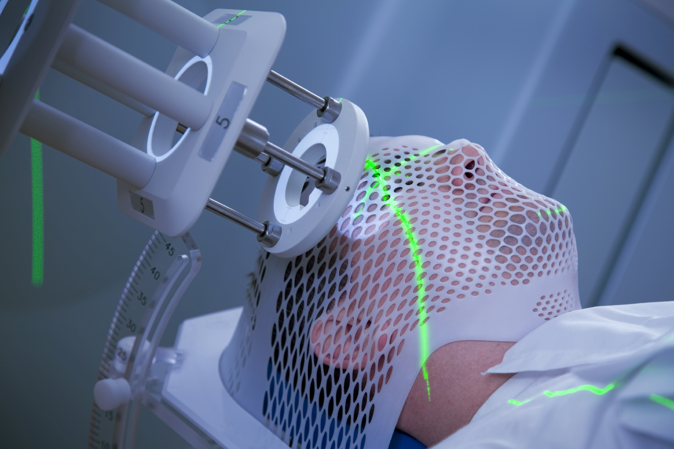
Figure 2: patient in a linear accelerator [10]
Next to the EBRT there are other radiation techniques that can be used for brain tumor treatment. These include:
- 3D conformal radiation therapy (3D-CRT): with MRI images the location of the tumor is mapped precisely; X-ray beams are directed to the tumor from different directions with low doses for each beam but an accumulation of radiation at the tumor arises
- intensity modulated radiation therapy (IMRT): This is an extension to the 3D therapy. Here the radiation is automatically delivered from different angles but the strength of each beam can be changed as well.
- conformal proton beam radiation therapy: similar to 3D-CRT, but protons are used instead of X-rays
- brachytherapy (internal radiotherapy): Here radiational material is directly inserted into or near to the tumor. It allows to treat the tumor with a high dose while keeping the effect on healthy brain tissue relatively small.
Radiation is used in the following cases:
- follow-up to surgery when microscopic tumor cells were left in the brain or the tumor was only partially resected
- for inoperable tumors
- for metastatic brain tumors
- for relieving symptoms (palliative radiation)
Risks and side effects of radiation include: fatigue, hair loss, ededma (brain swelling), blood clots and change in short-term memory.
Chemotherapy
In chemotherapy brain cancers are treated with different drugs to stop the tumor growth. This is done by either stopping them to reproduce or by initialising normal cell death (apoptosis). For the former method cytostatics are used. These interfer in a cell cycle to stop the reproduction of the cancer cell. In contrast, the latter one uses cytotoxics. Drugs can also be divided into cell-cycle specific and cell-cycle nonspecific drugs regarding whether they act at specific parts of the cell cycle.
| cytostatic | cytotoxic |
|---|---|
| Anti-Angiogenesis Inhibitors | Alkylating agents |
Drugs suppressing the repair damage mechanism of cells | Antimetabolites |
| Growth factor inhibitors | Hormones |
| Mitotic Inhibitors | |
| Steroids |
| cell-cycle specific | cell-cycle nonspecific |
|---|---|
| Hormones | Bevacizumab |
| Steroids | Cilengitide |
| Etoposide | Cisplatin |
| Hydroxyurea | Carmustine |
| Methotrexate | Lomustine |
| Procarbazine | Irinotecan |
| Temozolomide | Rapamycin |
| Vincristine |
Patients can be supplied with these drugs in two different ways. The systematic delivery works by injecting the drug into an artery, a vein or similar locations from which the drug reaches the bloodstream, crosses the blood brain barrier and enter the tumor cells. In contrast to that drugs are directly placed close to the tumor during local delivery. Thereby the drugs don't have to travel through the whole body and the concentration of the drug close to the tumor is increased.
Side effects and risks of chemotherapy can include vomiting, diarrhea, fatigue, hair loss, infertility brain swelling and cognitive problems.
Radiation therapy can be combined with chemotherapy by working simultaneously or being applied on after another.


Figure 3: A brief history of chemotherapy [11]
Auxiliary method: usage of steroids
Brain tumors and their treatments (especially radiation therapy) can cause a buildup of fluid, which is called edema. An edema can cause pressure on certain brain regions and therefore causes neurologic constraints.
The volume of edemas can be reduced by using steroids, the effect on the edema is temporarily, though. Steroids can improve neurological symptoms, increase the general feeling of well-being and appetite. As steroids are hormones, there are several side-effects when they are taken for longer time spans: weight gain, thinning of skin, upset stomach, muscle weakness, hiding a fever, mood swings, insomnia, pneumonia and increased blood sugar levels. [7]
Bibliography
[1] http://www.abta.org/brain-tumor-treatment/treatments/
[2] http://braintumor.org/brain-tumor-information/treatment-options/
[3] http://www.abta.org/secure/surgery.pdf
[4] http://www.abta.org/secure/conventional-radiation.pdf
[5] https://www.cancer.org/cancer/brain-spinal-cord-tumors-adults/treating/radiation-therapy.html
[6] http://www.abta.org/secure/chemotherapy.pdf
[7] http://www.abta.org/brain-tumor-treatment/treatments/steroids.html
[8] Surgical footage of removal of Glioblastoma Multiforme (GBM) Brain Tumor (brain surgery)
[9] https://www.cancer.gov/publications/dictionaries/cancer-terms?cdrid=46457
[10] http://dyingandgrief.com/brain-tumor-what-you-need-to-know
[11] ; A History of Cancer Chemotherapy; published in (2008); DOI:
Kommentar
Ardit Ramadani sagt:
31. Mai 2017Hi,
In general the wiki has a very good flow and the information is served appropriately. Without commenting much on the content, I would say that enough information is provided on the topics, however I personally would prefer a bit more details in Radiation therapy and Chemotherapy.
Furthermore, it is a matter of personal taste at the end (as we discussed in previous week), but I would avoid putting references in the title. I understand that the whole chapter is based on that reference, but, it does not show consistency between the references [6] and [7] for example. Chemotherapy is based completely on reference 6 and you have put it in the title, similarly Auxiliary method: usage of steroids is based on reference 7 and you have put it only at the end of paragraph.
Generally, I like the flow and the logic between sectioning of the pages. Keep it up with the good work!