Brain tumor symptoms
First of all, to know the symptoms of a brain tumor we need to know what a brain tumor is. With that being said, I would like to start with what means to have a brain tumor.
A brain tumor is the abnormal growth of a tissue on the brain which can disrupt important functions of the brain or even make them completely non-functional. [5]
The symptoms of brain tumor depend from tumor type, tumor size and it's location, which means that not all the symptoms listed below may occur to all the people with brain tumor. Most of the symptoms of brain tumor are very common and similar to other disease symptoms like :
 Headaches (usually worse in the morning)
- Nausea and vomiting
- Changes in speech, vision, or hearing (specific symptom related to brain tumor)
- Problems balancing or walking
- Changes in mood, personality, or ability to concentrate
- Problems with memory (specific symptom related to brain tumor)
- Muscle jerking or twitching (seizures or convulsions)
- Numbness or tingling in the arms or legs
Diagnosis
Since these symptoms can not immediately define if a person has brain tumor or not, the next phase is diagnosis. It is not easy to diagnose it either since there are a lot type of tumors (If you want to know more about tumor types you can click here!) and especially it is not easy at all to understand why a person can get a brain tumor unless it is genetic (which is a rare case) or a person has been exposed to radiation for a substantial period of his/her life. [2], [3], [7]
Listed below there are a number of analysis a person is asked to do before he/she is diagnosed with brain tumor. It is not mandatory for the patients to do all the analysis below, it all depends from their symptoms and results.
- Usually people with brain tumor have also other health problems so they are asked to do some routine analysis like blood analysis, electrolytes, liver function tests, and a blood coagulation (clotting) profile.
- If the person has mental-status change as the main symptom, blood or urine tests may be done to rule out drug use as a cause of such symptoms. [2], [3], [7]
Neurologic exam
The doctor checks the person's vision, hearing, alertness, muscle strength, coordination, and reflexes. He/She also examines eyes to look for swelling caused by a tumor pressing on the nerve that connects the eye and the brain. If responses are not normal, a brain scan will be ordered, or a patient will be referred to a neurologist or neurosurgical oncologist for more tests. [2], [7]
 source
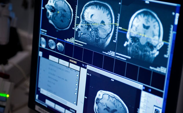
Scans AND Imaging Techniques
A scan is the first step to identify if a brain tumor is present, and to locate exactly where it is growing. A scan creates computerized images of the brain and spinal cord by examining it from different angles. Some scans use a contrast agent (or a dye) to allow the doctor to see the difference between normal and abnormal tissue. A patient may need more than one type of scan to diagnose a tumor, depending on its type and location. Commonly used scanning and imaging techniques:
Computed Axial Tomography(CT Scan)
CT scan is a computerized x-ray that can show a combination of soft tissue, bone, and blood vessels in 3D. This is often the first test a person will receive in an emergency room (i.e. after a seizure). A computerized tomography (CT) scan combines a series of X-ray images taken from different angles and uses computer processing to create cross-sectional images, or slices, of the bones, blood vessels and soft tissues inside your body. CT scan images provide more detailed information than plain X-rays do. A CT scan has many uses, but is particularly well-suited to quickly examine people who may have internal injuries from car accidents or other types of trauma. A CT scan can be used to visualize nearly all parts of the body and is used to diagnose disease or injury as well as to plan medical, surgical or radiation treatment. [2], [3], [6], [7], [10],

Left: Arrows indicate a collection of blood between the skull and the outer covering of the brain (epidural hematoma) that's compressing the frontal lobe. Right: Contrast material injected into a vein during this CT scan of the head highlights tumors in both sides of the brain.
Magnetic Resonance Imaging (MRI)
MRI can create clear and detailed three-dimensional images of a brain tumor. An MRI is not often used with people who have a pace maker or other metal device. Has a higher sensitivity to detect the presence of a tumor and also characteristics of a tumor, if there is one. Magnetic resonance imaging (MRI) is a technique that uses a magnetic field and radio waves to create detailed images of the organs and tissues within your body. [12]
Magnetic Resonance Spectroscopy (MRI Spect or MRS)
MRI Spect measures the levels of metabolites in the body.
An MRS can detect irregular patterns of activity to help diagnose the type of tumor, evaluate its response to therapies, or determine aggressiveness of a tumor.
Perfusion MRI
Perfusion MRI examines the flow of blood into the tissues to help assess the grade/aggressiveness of tumors and differentiate a recurrent tumor from dead tumor tissue.
Functional MRI (fMRI)
FMRI tracks the use of oxygen and blood flow in the brain as patients perform tasks.
An fMRI can identify the motor, sensory, visual and language centers of the brain which helps your doctor carefully plan for surgery. The eloquent brain can be identified using functional MR (fMR) imaging for the gray matter and diffusion tensor (DT) imaging for the white matter. fMR imaging and DT imaging are especially important for patients with tumors near the important motor and language centers of the brain, where the normal anatomic references may be distorted by the tumor and associated edema. It may be used to examine the brain’s anatomy, determine which parts of the brain are handling critical functions, evaluate the effects of stroke or disease, or guide brain treatment. fMRI may detect abnormalities within the brain that cannot be found with other imaging techniques. [14], [15] Figure 1.
 Different relations between the eloquent brain areas and the brain tumors as shown by fMRI. (A) PMA for the hand (white arrow) is more than 2 cm from the tumor (grey arrow) (B) PMA for the right foot (white arrow) is 1–2 cm from the tumor (grey arrow). (C) PMA for the right hand (white arrow) is less than 1 cm from the tumor (grey arrow). (D) Broca's area is totally infiltrated by the tumor (grey arrow).
Positron Emission Tomography (PET)
PET scan uses a radioactive substance to visualize hypermetabolic activity such as with malignant cells, or abnormalities from a tumor or scar tissue.
A positron emission tomography (PET) scan is an imaging test that helps reveal how your tissues and organs are functioning. A PET scan uses a radioactive drug (tracer) to show this activity. The tracer may be injected, swallowed or inhaled, depending on which organ or tissue is being studied by the PET scan. The tracer collects in areas of your body that have higher levels of chemical activity, which often correspond to areas of disease. On a PET scan, these areas show up as bright spots. A PET scan is useful in revealing or evaluating several conditions, including some cancers, heart disease and brain disorders. PET is also used during brain mapping procedures. The pictures from a PET scan provide information different from that uncovered by other types of scans, such as computerized tomography (CT) or magnetic resonance imaging (MRI). A PET scan or a combined CT-PET scan enables your doctor to better diagnose your condition. [16]
Spinal tap
The doctor may remove a sample of cerebrospinal fluid (the fluid that fills the spaces in and around the brain and spinal cord). This procedure is performed with local anesthesia. He/She uses a long, thin needle to remove fluid from the lower part of the spinal column. A spinal tap takes about 30 minutes. The patient must lie flat for several hours afterward to keep from getting a headache. A laboratory checks the fluid for cancer cells or other signs of problems. Spinal tap (also called a lumbar puncture), uses a special needle placed into the lower back to measure pressure in the spinal canal and brain and determine if there is an infection or tumor cells. [2]


Angiogram
Dye injected into the bloodstream makes blood vessels in the brain show up on an x-ray. If a tumor is present, the x-ray may show the tumor or blood vessels that are feeding into the tumor.
Cerebral angiogram showing brain AVM [18]
Biopsy

The removal of tissue to look for tumor cells is called a biopsy. A pathologist looks at the cells under a microscope to check for abnormal cells. A biopsy can show cancer, tissue changes that may lead to cancer, and other conditions. A biopsy is the only sure way to diagnose a brain tumor, learn what grade it is, and plan treatment. Surgeons can obtain tissue to look for tumor cells in two ways: [2], [3]
Biopsy at the same time as treatment
The surgeon takes a tissue sample when you have surgery to remove part or all of the tumor. This is also called open biopsy which is performed during a craniotomy. Craniotomy is part of tumor treatments. (To learn more about tumor treatments click here)Stereotactic biopsy
You may get local or general anesthesia and wear a rigid head frame for this procedure. The surgeon makes a small incision in the scalp and drills a small hole (a burr hole) into the skull. CT or MRI is used to guide the needle through the burr hole to the location of the tumor. The surgeon withdraws a sample of tissue with the needle. A needle biopsy may be used when a tumor is deep inside the brain or in a part of the brain that can't be operated.
Myelogram
The doctor may recommend a myelogram to find out if the tumor has spread to the spinal fluid, other parts of the brain, or the spinal cord. A myelogram uses a dye injected into the CSF that surrounds the spinal cord. The dye shows up on an x-ray and can outline the spinal cord to help the doctor look for a tumor. This is rarely done; a lumbar puncture is more common. [7]Molecular testing of the tumor
Your doctor may recommend running laboratory tests on a tumor sample to identify specific genes, proteins, and other factors, such as tumor markers, unique to the tumor. Some biomarkers may help doctors determine a patient’s prognosis. Researchers are examining biomarkers to find ways to diagnose a brain tumor before symptoms begin. Ultimately, results of these tests may help decide whether your treatment options include a type of treatment called targeted therapy. [7]
Bibliography:
- http://abc2.org/guidance/brain-cancer-facts/symptoms
- http://abc2.org/guidance/brain-cancer-facts/diagnosis
- http://abc2.org/sites/default/files/Frankly%20Speaking%20About%20Cancer_Brain%20Tumors_0.pdf
- http://www.cancer.net/cancer-types/brain-tumor/symptoms-and-signs
- http://www.emedicinehealth.com/brain_cancer/article_em.htm
- http://www.emedicinehealth.com/brain_cancer/page6_em.htm#what_exams_and_tests_diagnose_brain_cancer
- http://www.cancer.net/cancer-types/brain-tumor/diagnosis
- http://www.abta.org/brain-tumor-information/?referrer=http://www.abta.org/brain-tumor-treatment/newly-diagnosed/ (General information taken)
- http://www.ourhealthpage.com/what-is-brain-cancer/#prettyPhoto
- http://www.mayoclinic.org/tests-procedures/ct-scan/basics/definition/prc-20014610
- http://www.mayoclinic.org/tests-procedures/ct-scan/multimedia/ct-scan-images-of-the-brain/img-20008347
- http://www.mayoclinic.org/tests-procedures/mri/home/ovc-20235698
- https://www.ncbi.nlm.nih.gov/pubmed/20708553
- https://www.radiologyinfo.org/en/info.cfm?pg=fmribrain
- http://www.sciencedirect.com/science/article/pii/S0378603X11000349
- http://www.mayoclinic.org/tests-procedures/pet-scan/basics/
- https://www.google.de/search?q=pet+brain+images:
- http://www.mayoclinic.org/diseases-conditions/brain-avm/multimedia/brain-avm/img-20006461
3 Kommentare
Unbekannter Benutzer (ga67yur) sagt:
16. Mai 2017Nice wiki entry. I like your description of how the techniques work and also what they show/why they are useful.
Maybe you could add links from the bibliography entries to your text and images so it's easier to track which source you used for which part, if possible.
And I noticed that the "you can click here" part at the beginning of your diagnosis part is not working.
Apart from that great work!
Unbekannter Benutzer (ga94leq) sagt:
16. Mai 2017Unbekannter Benutzer (ga67yur) thank you once again I just fixed the link so it works now. And as I already said I forgot to link the images so I am going to do that to. I am really glad you think so
Unbekannter Benutzer (ga39tec) sagt:
17. Mai 2017In addition to Christine's comment, maybe you could link the tumor symptoms to brain regions. But the tumor symptoms part is a good overview nonetheless!