Overview
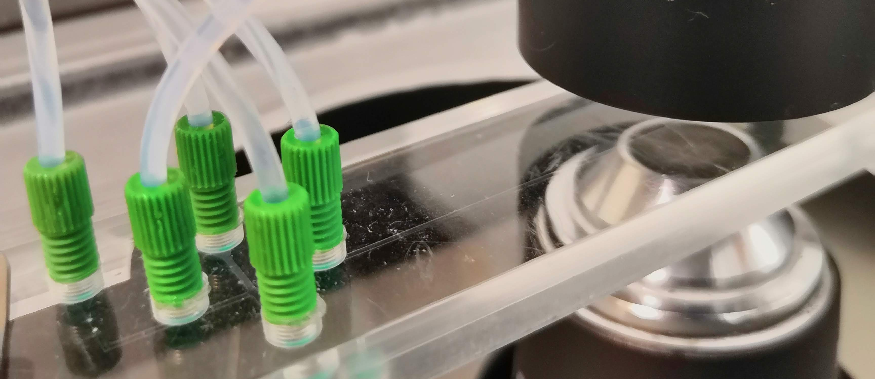
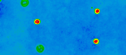
CellFace aims to establish digital, holographic flow cytometry as a label-free platform technology for cellular diagnostics. We are an interdisciplinary project of TU Munich coordinated by the Chair for Biomedical Electronics and the Chair for Data Processing in cooperation with several departments of the University Hospital Klinikum rechts der Isar.
Motivation
Blood delivers a variety of insights into health conditions of an organism, especially in the field of in-vitro diagnostics. Therefore, hematological analysis such as complete blood count (CBC) represent a high share of laboratory tests in the health care sector. Despite the availability of largely automated analyzers, the gold standard for the routine diagnosis of hematological disorders is the tedious Giemsa stained blood smear [1] which suffers from inter-observer variations. Further drawbacks of current approaches are a high effort for sample preparation, material expenses, a long processing time or missing clinical relevance [2]. To overcome the mentioned issues and specially to reduce time-consuming manual intervention, the CellFace project was created. Flow cytometry in combination with digital holographic microscopy (DHM) [3] enables researchers to record 3D images of each single cell, while sustaining a high throughput stream of blood cells. The overall goal is to establish this label- and reagent-free approach as a platform technology for automated blood cell diagnosis [4]. The best methodologies to analyze the recorded phase images are subject to research. Therefore, this work will investigate classical and modern techniques in the field of computer vision and machine learning on their suitability for interpreting the images and the contained information.
Scientific and Technical Goals
We develop a novel in vitro diagnostic method for cell analysis and biomarkers which are not accessible today for clinical routine. With access to clinical samples, a highly accurate sample presentation and imaging solution, we can offer AI-induced data exploration and interpretations of volatile biomarker panels.
A tiny drop of blood gives us a detailed picture of patient health and lets us support clinicians in their decision-making. Our label-free technology enables us to provide insights in different health aspects varying from cardiovascular risk assessment to leukemia detection and all that within just a few minutes.
Publications
| Year | Title | Link | Mediatum / FIS |
|---|---|---|---|
2024 | Unsupervised high-throughput segmentation of cells and cell nuclei in quantitative phase images | https://arxiv.org/abs/2311.14639 | FIS |
2024 | Immunothrombosis of acute care patients quantified with phase imaging flow cytometry | SPIE | |
2023 | Platelet aggregates detected using quantitative phase imaging associate with COVID-19 severity | Nature | FIS |
| 2023 | Explainable Artificial Intelligence for Cytological Image Analysis | AIME 2023 | FIS |
| 2023 | Composition Counts: A Machine Learning View on Immunothrombosis using Quantitative Phase Imaging | ML4HC 2023 | FIS |
| 2023 | Measurement of Platelet Aggregation in Ageing Samples and After in-Vitro Activation | BIOIMG 2023 | MediaTUM |
| 2023 | Towards Interpretable Classification of Leukocytes based on Deep Learning | ICML 2023 | MediaTUM |
| 2023 | Explainable Feature Learning with Variational Autoencoders for Holographic Image Analysis | BIOIMG 2023 | MediaTUM |
| 2023 | Rethinking Usability Heuristics for Modern Biomedical Interfaces | ACHI 2023 | MediaTUM |
| 2023 | Platelet micro-aggregate diagnostics with quantitative phase microscopy | SPIE | |
| 2022 | Outlier Detection using Self-Organizing Maps for Automated Blood Cell Analysis | ICML 2022 | MediaTUM |
| 2022 | A Comparison of Uncertainty Quantification Methods for Active Learning in Image Classification | IJCNN 2022 | MediaTUM |
| 2022 | Label-free Digital Holographic Microscopy to Characterize Inter- and Intratumoral Heterogeneity in Pancreatic Cancer | Mo1219 | MediaTUM |
| 2020 | Impact of sample preparation on holographic imaging of leukocytes | SPIE | |
| 2020 | Morphology–Here I Come Again | Cytometry Part A | MediaTUM |
| 2020 | Autoencoder Features for Differentiation of Leukocytes based on Digital Holographic Microscopy (DHM) | EUROCAST 2019 | FIS |
Our Partners
Latest Activity
Contributors
References
- J. J. Barcia, “The giemsa stain: Its history and applications,” International Journal of Surgical Pathology, vol. 15, no. 3, pp. 292–296, 2007.
- A. Filby, “Sample preparation for flow cytometry benefits from some lateral thinking,” Cytometry Part A: the journal of the International Society for Analytical Cytology, vol. 89, pp. 1054–1056, 2016.
- Y. Jo, H. Cho, S. Yun Lee, G. Choi, G. Kim, H.-s. Min, and Y. Park, “Quantitative phase imaging and artificial intelligence: A review,” IEEE Journal of Selected Topics in Quantum Electronics, vol. 25, pp. 1–14, 2018.
- Z. El-Schich, S. Kamlund, B. Janicke, K. Alm, and A. Wingren, “Holography: The usefulness of digital holographic microscopy for clinical diagnostics,” in Holographic Materials and Optical Systems, pp. 319– 333, Intech, 2017.
- S. Prieto, A. Powless, J. W. Boice, S. Sharma, and T. Muldoon, “Proflavine hemisulfate as a fluorescent contrast agent for point-of-care cytology,” PLoS ONE, vol. 10, p. e0125598, 2015.
- M. Ugele, M. Weniger, M. Stanzel, M. Bassler, S. W. Krause, O. Friedrich, O. Hayden, and L. Richter, “Labelfree high-throughput leukemia detection by holographic microscopy,” Advanced Science, vol. 1800761, 2018.
- M. Rinehart, H. Sang Park, and A. Wax, “Influence of defocus on quantitative analysis of microscopic objects and individual cells with digital holography,” Biomedical Optics Express, vol. 6, no. 6, pp. 2067–2075, 2015.
- Y. Li, B. Cornelis, A. Dusa, G. Vanmeerbeeck, D. Vercruysse, E. Sohn, K. Blaszkiewicz, D. Prodanov, P. Schelkens, and L. Lagae, “Accurate label-free 3-part leukocyte recognition with single cell lens-free imaging flow cytometry,” Computers in biology and medicine, vol. 96, pp. 147–156, 2018.
- D. Vercruysse, A. Dusa, R. Stahl, G. Vanmeerbeeck, K. de Wijs, C. Liu, D. Prodanov, P. Peumans, and L. Lagae, “Three-part differential of unlabeled leukocytes with a compact lens-free imaging flow cytometer,” Nature biotechnology, vol. 15, pp. 1123–1132, 2013.
- E.-A. D. Amir, K. L. Davis, M. Tadmor, E. Simonds, J. Levine, S. Bendall, D. Shenfeld, S. Krishnaswamy, G. Nolan, and D. Pe’er, “visne enables visualization of high dimensional single-cell data and reveals phenotypic heterogeneity of leukemia,” Lab on a Chip, vol. 31, no. 6, pp. 545–52, 2013.
- D. Roitshtain, L. Wolbromsky, E. Bal, H. Greenspan, L. L. Satterwhite, and N. T. Shaked, “Quantitative phase microscopy spatial signatures of cancer cells,” Cytometry Part A: the journal of the International Society for Analytical Cytology, vol. 91, pp. 482–493, 2017.
- J. Yoon, Y. Jo, M.-h. Kim, K. Kim, S. Lee, S.-J. Kang, and Y. Park, “Identification of non-activated lymphocytes using three-dimensional refractive index tomography and machine learning,” Scientific reports, vol. 7, no. 1, p. 6654, 2017.


