In this page, anatomy of the brain will be explain, including the histology of the brain, its structure and components. Furthermore, functions related to each part and component of the brain will be briefly discussed.
Histology of the Brain
In the cellular level, we can identify two main types of cells within the brain: Neurons and Neuroglia.
- Neurons: They are specialised cells that carry electrical impulses and perform all the communication and processing in the brain. They have the following structure:
- Dendrites: Extend from the cell body. Short with many branches. They receive the neural input form other neurons via synapsis and transmit it to the perikaryon.
Cell body or perikaryon.
Axon: single nerve finer which transmits the impulses coming from the perikaryon to the distal end. The length can vary from a few microns to one meter.
Moreover, according to the number of structures extending from the cell body, a neuron can be unipolar (one structure), bipolar (one dendrite and one axon) or multipolar (one axon and many dendrites). According to their function and location, we can distinguish three types of neurons:- Sensory neurons: They enter the brain from the peripheral nervous system and deliver information about the condition to the body and the surroundings.
Interneurons: integrate and process the information delivered by the sensory neurons. Fully located in the brain.
Motor neurons: Receive the signals from the interneurons and carry them to the muscles and glands.

Structure of a neuron [1]
Neuroglia: They support and protect the neurons. They can be classified into four types. This type of cells are present within the central and peripheral nervous system, however we will only refer to those present in the first one, as is where the brain is located.
Astrocytes: they protect the neurons by filtering nutrients out of the blood and preventing chemicals and pathogens from leaving the capillaries of the brain.
Oligodendrocytes: They warp the axis of the neurons to produce the insulation known as myelin, increasing communication speed between neurons.
Microglia: their function is quite similar to the white blood cells by attacking and destroying the pathogens that enter the brain.
Ependymal : line the capillaries of the chord plexuses and filter blood plasma to produce the cerebrospinal fluid (CSF)

Scheme of the interaction between the neurons and the glia cells in the central nervous system [2]
Both types of brain cells, in combination, form two types of brain tissue: Grey matter and white matter:
Grey matter: It is mostly made of neural perikaryon and glia cells. Non-myelinated (dendritic) processes take place here. They are the areas of nerve connections and processing, i.e. where synapsis takes place.
White matter: made mostly of myelinated axons and oligodendrocytes. It connects regions of grey matter to each other and to the rest of the body. The white matter acts like the information highway of the brain.

Slice of brain differentiating the main structures and tissues. [3]
Structure of the Brain
Based on embryonic development, brain is usually divided into three main regions:
- Forebrain or prosencephalon
- Midbrain or mesencephalon
- Hindbrain or rhombencephalon

Scheme of the location of the brain regions in sagittal view. [4]
Hindbrain
The Hindbrain contains the brainstem and the cerebellum.
- Brainstem: It connects the brain to the spinal cord. It is the most inferior part of the brain. Most of the basic survival functions are controlled in this area. The brainstem is itself divided into three regions: the medulla oblongata and the pons. All of these regions contain a net-like structure composed of mixed grey and white matter. This structure known as reticular formation controls the muscle tone in the body and acts as a switch between the consciousness and sleep in the brain.
- The medulla oblongata is a cylindrical mass of nervous tissue that connects the spinal cord on its inferior border and the pons on its superior one. It mostly contains white matter that carries nerve signals ascending to the brain and descending to the spinal cord. There are also several regions of gray matter that process involuntary body functions related to homeostasis such us: blood pressure and oxygen levels monitoring and heart rate regulation in the cardiovascular center of the medulla; breathing rate in the rhythmicity center, and some reflexes such as vomiting, sneezing, coughing and swallowing.
- The pons is found superior to the medulla oblongata, inferior to the midbrain and anterior to the cerebellum. Together with this last one forms the mesencephalon. The pons acts as a bridge for nerve signals travelling from and to the cerebellum, and carries signals between the superior regions of the brain and the medulla, and the spinal cord.
- The medulla oblongata is a cylindrical mass of nervous tissue that connects the spinal cord on its inferior border and the pons on its superior one. It mostly contains white matter that carries nerve signals ascending to the brain and descending to the spinal cord. There are also several regions of gray matter that process involuntary body functions related to homeostasis such us: blood pressure and oxygen levels monitoring and heart rate regulation in the cardiovascular center of the medulla; breathing rate in the rhythmicity center, and some reflexes such as vomiting, sneezing, coughing and swallowing.
- Cerebellum: It’s a wrinkled, hemispherical region of the brain that is located posterior to the brainstem and inferior to the cerebrum. The outer layer is called cerebellar cortex, and it is made of tightly folded grey matter that provides the processing power of the cerebellum. After the cerebellar cortex there is a tree-shaper layer of white matter called arbor vitae, which connects the processing regions of the cerebellar cortex to the rest of the brain and body.
The main function of the cerebellum is help to control motor functions such as balance, posture and coordination of complex muscle activities. It received sensory inputs from the muscles and joins of the body and uses the information to keep the body balance and to maintain posture. It also controls the timing and finesse of complex motor actions such as walking, writing and speech.


Sagittal view of the brain. For left to right: Hindbrain, Medulla Oblongata, Pons and Cerebellum [5]
Midbrain
It is the most superior regions of the brainstem. It is found between the pons and the diencephalon. It can be subdivided into two regions: the tectum and the cerebral peduncles.
- Tectum: Is the posterior region of the midbrain and it contains relays for reflexes that involve auditory and visual information. Also reflexes such as pupillary, accommodation and startle reflexes are relayed through this region.
- Cerebral Peduncles: They form the anterior part of the midbrain, and they contain many nerve tracts and the substantia nigra. The nerves passing through the cerebral peduncles connect regions of the cerebrum (basal ganglia) and thalamus to the spinal cord and lower regions of the brainstem. The substatia nigra is a region of neurons that contain dark melanin that is involved in the inhibition of movement. Parkinson’s disease is related to the degeneration of this area. The substatia nigra is made up of two anatomically and functionally distinct parts: substantia nigra pars compacta and substantia nigra pars reticulata. Neurons in the pars compacta are more densely packed together than in the pars reticulata. The dopamine neurons are mostly found in the pars compacta, while in the pars reticulata we can find neurons containing GABA, which is a primary inhibitor neurotransmisor.

Forebrain
It can be divided into diencephalon and cerebrum.
- Diencephalon: It is located superior and anterior to the midbrain. These are the main regions: thalamus, hypothalamus and pineal gland.
- Thalamus: It’s formed by a pair of oval masses of grey matter inferior to the lateral ventricles and surrounding the third ventricle. The sensory neurons that enter the brain from the peripheral nervous system form relays with neurons in the thalamus that continue on to the cerebral cortex. Therefore, the thalamus acts like a switchboard operator of the brain by routing sensory inputs to the correct regions of the cerebral cortex. It has also an important role in learning by routing sensory information into processing and memory centres of the cerebrum.
- Hypothalamus: It is located inferior to the thalamus and superior to the pituitary gland. It acts as the brain’s control center for body temperature, hunger, thirst, blood pressure, heart rate and hormones production. In response to the changes in the body’s condition detected by the sensory receptors, the hypothalamus sends signals to glans, smooth muscles and heart to counteract these changes. It also controls directly the pituitary gland by producing hormones. Some of them are produced in the hypothalamus itself and stored in the pituitary gland (oxytocin and antidiuretic hormone), and others (releasing and inhibiting hormones) are secreted directly into the blood stream in order to stimulate or inhibit hormone production in the anterior pituitary gland.
- Thalamus: It’s formed by a pair of oval masses of grey matter inferior to the lateral ventricles and surrounding the third ventricle. The sensory neurons that enter the brain from the peripheral nervous system form relays with neurons in the thalamus that continue on to the cerebral cortex. Therefore, the thalamus acts like a switchboard operator of the brain by routing sensory inputs to the correct regions of the cerebral cortex. It has also an important role in learning by routing sensory information into processing and memory centres of the cerebrum.
/about/diencephalon-5755b3c23df78c9b467a408c.jpg)
Image showing the location of the structures of the diencephalon plus the cerebellum [7]
- Pineal gland: It is a small gland located posterior to the thalamus in a subregion called epithalamus. It produces the melatonin hormone. In the dark, it secretes melatonin which has a sedative effect and helps to induce sleep. The production rate of melanin is decreased with age.
- Pineal gland: It is a small gland located posterior to the thalamus in a subregion called epithalamus. It produces the melatonin hormone. In the dark, it secretes melatonin which has a sedative effect and helps to induce sleep. The production rate of melanin is decreased with age.

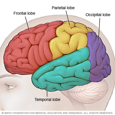
- Cerebrum: It is the largest region of the brain. It controls higher brain functions such as language, logic, reasoning and creativity. It surrounds the diencephalon and is is located superior to the cerebellum and brainstem. It is divided into left and right hemisphere by the longitudinal fissure, that runs midsagitally down to the center of the cerebrum. Each hemisphere can be divided into 4 lobes: frontal, parietal, temporal and occipital, named by the skull bones that cover them .The hemispheres are organised into funcional areas: motor, sensory and association. The right side of the body is under control of the left hemisphere and the left side under the right hemisphere, this characteristic is called contralateral control. The functions are lateralised, i.e. each hemisphere has unique functions, which arise from concerted activity. The hemispheres are connected via the corpus callosum, located above the thalamus, under the cortex. It is responsible for transmitting the neural messages between both hemispheres.

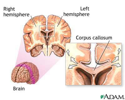
From left to right. Location of the Cerebrum in a sagittal view of the brain [5]. Location of the corpus callosum in an axial view [9]. Lobes of the Cerebrum in sagittal view [10]
The surface is a convoluted layer of grey matter known as cerebral cortex, where most of the processing of the cerebrum takes place. These convolutions separate and divide the lobes from both hemispheres. There are three types of these surface convolutions:- Gyrus: A ridge on the surface.
- Sulcus: A groove.
- Fissure: A deep groove.
- Deep in the cerebrum white matter, there are several regions of grey matter that form the basal nuclei and the limbic system.
- Basal nuclei or ganglia: They work together with the substatia nigra to regulate and control muscle movements. Often the substantia nigra is included into the basal ganglia, as well as other structures of the diencephalon such as the subthalamic nucleus. It helps to control muscle tone, posture and subsconcious skeleton muscle. In the brain they include: caudamen, putamen and globus pallidus. They receive information from the cerebral cortex, mostly through the caudamen and putamen, it is processed and sent back to the cortex via the thalamus. To know more about the basal nuclei, click here
- Limbic system: Set of structures located on top of the brain stem and buried under the cortex. It includes the hippocampus and the amygdala. Helps the body to react to emergency and highly emotional situations with fast and almost involuntary actions.
- Hippocampus: It is located in both temporal lobes. It is the primary output node of the limbic system. It assists with the storage of long term memories and it's also responsible for the memory of location of objects and people. It acts as a memory indexer, sending the memories to the right part of the hemisphere for storage.
- Amygdala: Almond mass shape of nuclei involved in hormonal secretion, emotional responses and memory. It is responsible for fear conditioning and the associative learning process, by which humans learn to fear something. It is located adjacent to the hippocampus.
- Basal nuclei or ganglia: They work together with the substatia nigra to regulate and control muscle movements. Often the substantia nigra is included into the basal ganglia, as well as other structures of the diencephalon such as the subthalamic nucleus. It helps to control muscle tone, posture and subsconcious skeleton muscle. In the brain they include: caudamen, putamen and globus pallidus. They receive information from the cerebral cortex, mostly through the caudamen and putamen, it is processed and sent back to the cortex via the thalamus. To know more about the basal nuclei, click here

Basal nuclei in axial view: caudate nucleus, putamen, globus palidus, subthalamic nucleus and substantia nigra. [11]


Protection of the Brain
Between the skull and the brain, there are three layers of tissue that surround and protect the brain and the spinal cord. These layers are called meninges and are called: dura mater, arachnoid mater and pia mater.
- Dura mater: It’s the outermost layer of the meninges. It is a dense irregular connective tissue made of tough collagen fibres. It forms a pocket around the brain and the spinal cord to hold the CSF and prevent mechanical damage to the soft tissue underneath.
- Arachnoid mater: It is much more thinner and delicate than the dura mater. It contains thin fibres resembling to a spider web, that connect the dura mater with the pia mater. Beneath this layer is a flus-filled region called subarachnoid space.
- Pia mater: I t is the innermost layer of the meninges. It is placed directly ont he surface of the brain and the spinal cord. It provides oxygen and nutrients to the nervous tissue of the brain. It also helps regulating the flow of materials from the bloodstream and CSF to the nervous system.

Structures located between the skull and the cerebral cortex [14]
Furthermore, surrounding the brain and the spinal cord there is fluid called Cerebrospinal fluid (CSF), and together with the meninges, collaborate in the protection of the brain. It fills the subarachnoid space and exerts pressure on the outside of the brain and spinal cord. This pressure acts as stabiliser and shock absorber for the brain and spinal cord, as they float within the hollow spaces of the skull and vertebrae. Inside the brain, the CSF fills small cavities called ventricles, that expand under the pressure of the CSF to lift and inflate the soft brain tissue.
The CSF is produced in the brain by the choroid plexuses. It is released into the arachnoid space and it contains glucose, oxygen and ions, which will be distributed throughout the nervous system. It also transports waste products away. After circulating around the brain and spinal cord, it enters small structures called arachnoid vilis, where it is reabsorbed into the bloodstream.
In order to maintain a normal brain function, the neural environment must be preserved within a narrow homeostatic range. This requires a tight regulation of cells, molecules and bones between the blood and the brain. Such regulation is maintained by an anatomical and physiological barrier called Blood-Brain Barrier (BBB). This barrier is made of highly specialised endothelial cells (EC) that line brain capillaries and transduce signals from the vascular system and from the brain. The BBB is located outer the CSF and the CSF picks up the essential molecules that diffuse via the junction gaps of the BBB from the blood stream.
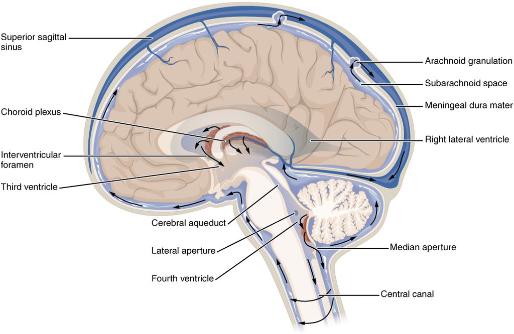
Circulation of the CSF in the brain [15]

Section through a brain blood vessel obtained with confocal light microscopy. The dark area is the lumen of the vessel. Surrounding the blood vessel are glial cells (green), which provide support to neurons (red). [16]
Bibliography
[1] http://ehumanbiofield.wikispaces.com/Neurons
[2] https://cnx.org/contents/c9j4p0aj@3/Neurons-and-Glial-Cells
[4] https://phys-mic-vic-ash.wikispaces.com/file/view/brain-divisions.gif/301425706/brain-divisions.gif
[5] http://www.innerbody.com/image/nerv02.html
[7] https://www.thoughtco.com/diencephalon-anatomy-373220
[8] http://upliftconnect.com/detoxify-your-pineal-gland/
[9] https://psychlopedia.wikispaces.com/Corpus+Callosum
[10] http://www.mayoclinic.org/brain-lobes/img-20008887
[11] https://kin450-neurophysiology.wikispaces.com/Basal+Ganglia+II
[12] http://www.brainfacts.org/brain-basics/neuroanatomy/articles/2014/blood-brain-barrier
[13] http://www.sciencedirect.com/science/article/pii/S0969996103002833
[14] http://missinglink.ucsf.edu/lm/ids_104_cns_injury/response%20_to_injury/meninges.htm
[15] https://en.wikipedia.org/wiki/Cerebrospinal_fluid
[16] http://micro-scopic.tumblr.com/post/14105906627/blood-brain-barrier-confocal-light-micrograph-of
[17] http://www.histology.leeds.ac.uk/tissue_types/nerves/Nerve_neuron.php
[18] http://histology.medicine.umich.edu/resources/central-nervous-system#ii-neurons-slide-65-glial-cells
[19] http://www.neuroscientificallychallenged.com/blog/know-your-brain-substantia-nigra
[20] http://brainmadesimple.com/hippocampus.html
[21] http://brainmadesimple.com/amygdala.html
[22] http://brainmadesimple.com/corpus-callosum.html
[23] https://www.ncbi.nlm.nih.gov/pubmed/15207256