This part deals with different methods for brain tumor diagnosis. First we describe the neurological examination. Next we talk about invasive techniques and different analysis of the resulting samples. Lastly we introduce different medical imaging techniques.
Neurological examination
A neurological examination serves to evaluate the patients nervous system and helps to locate tumors in the brain. It can be performed without harming the patient.
During the examination the physican looks at the following aspects:
- Mental status: Here the awareness of the patient and his/her interaction with the environment is checked. This is done by pure observation during normal interactions and conversations.
- Motor system: Motor functions (activation and coordination of muscles and limbs) are checked by letting the patient push and pull with arms and legs against the physician. Balance can be analyzed by slightly pushing the patient while he/she has her eyes closed or by simply looking at the patients walk.
- Sensory system: These tests analyze the patients ability to feel by touching the patient at different parts of the body (legs, arms) and identifying the sensations (hot/cold sharp/dull).
- Reflexes: Here reflexes are examined by using a reflex hammer and examing the movement in the patients body caused by the reflex hammer.
- Cranial nerves (CNs): The brain has 12 of them, all of which are tested to evaluate different functions of the brain.
- Coordination: This is tested by letting the patient walk down a line or tapping his fingers quickly.
For the examination the physician can use the following tools: stethoscope, ophthalmoscope, reflex hammer, tuning fork, sterile safety pins.
For more details on the neurological examination, please check both links provided below.
 Figure 1: example template for a neurological exam [18]
Biopsy
Here, a small amount of tumor tissue is removed surgically and sent to the lab afterwards to be evaluated by a patheologist. This can be done as an individual surgery or as part of the surgery for tumor removal. For brain tumors, three types of biopsy are common:
- Needle biopsy: A small hole in the skull is opened through which the surgeon removes tumor tissue with a needle.
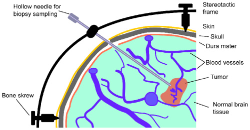
- Stereotactic biopsy: This procedure also uses a needle to remove tumor tissue, but the tools are guided by a computer system that uses CT or MRI scans to provide information about the exact location of the tumor.
- Open biopsy: The tumor sample is extracted during surgery while the tumor is exposed.
Laboratory Tests
There are different test in this group. While some are used to check for the existance of a tumor, others can indicate the impact of treatments and others show the growth of a tumor. The most common tests are described below:
- Lumbar Puncture (Spinal Tap): In this test a sample of the cerebrospinal fluid (CSF) is taken by a needle biopsy and is examined to see whether tumor cells, tumor markers, blood, infections or proteins are present. The resulting information can be used to diagnose primary CNS lymphoma and pineal or meningeal region tumors. Furthermore the composition of the sample can indicate whether a tumor has spread or not.

- Myelogram: Here, a special dye (contrast medium) is inserted into the patients spinal cord through a small needle. By taking X-Ray images afterwards, spinal tumors can be analyzed and located. Nowadays the X-Rays are often replaced with MRI.
- Evoked-potentials: The electrical activity of nerves, which are stimulated, is measured by using an EEG. Results can help to diagnose a vestibular schwannoma and to check neurological functions during brain surgery.
- Audiometry: This is a hearing test that is helpful to diagnose a cerebelloponte angel tumor.
- Endocrine Evaluation: Here the measurements of hormone levels in blood and urine samples are used diagnose pituitary and hypothalamic tumors.
- Perimetry: By measuring the size of the visual fields it is possible to reason about tumors in the area of the optic chiasm.
- Biomarkers: Bodily fluids contain tiny bits of genetic material from brain tumor cells. By monitoring them, the growth of the tumor and the effect of different treatment therapies can be analyzed. However this technique is still part of research and not commonly used in practice.
DNA profiling
This lab test is used to determine specific features of the patients DNA. A blood sample from the patient is analyzed and the resulting detailed information about the tumor can be used to define a personalized treatment plan. Analyzed features include gene mutations and other changes in the genetic field due to which one can draw conclusions regarding the origin and growth of the tumor and its response to different treament drugs.
Brain scans
Medical imaging in general is used to get detailed infomation about structures and functions of the body. Brain scans techniques can be divided into anatomical and functional imaging. Furthermore the scans can be contrast-enhanced or not. By using a contrast medium, particular parts of the brain (tissue, tumor, blood vessels, etc.) can be seen more clearly.
Anatomical imaging
Imaging techniques in this group show the motionless anatomy of the brain.
Functional imaging
In general these techniques are used for imaging of the functions and motions inside the brain. So bloodflow, oxygen saturation and vessel formation can be seen here among others.
Computed tomography (CT)
By moving the X-Ray beam in a circle around the brain, many images of the brain are taken from different angles. These so called slides are then joint to form a two or three dimensional model of the brain. Due to the rotation of the beam, the CT is more detailed than normal X-Ray. A CT scan can reveal information about the brain tissue and brain structures which can be used to examine location and size of the tumor as well as to evaluate effects of treatment methods. [9]
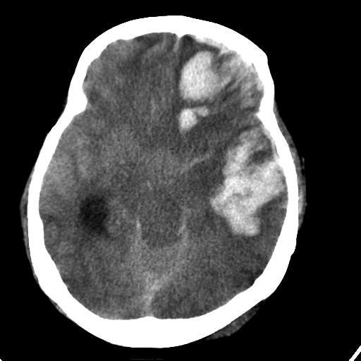
Figure 4: Axial CT showing an intra cranial hemorrhage [21]
Magnetic Resonance Imaging (MRI)
This technique works similar to CT but instead of X-Rays, magnetic fields and radio waves are used. Then from the resulting image slices, a two or three dimensional model can be created. It is used for studying organs or soft tissue, because the difference between normal and abnormal soft tissue can be seen better than with CT.
MRIs can be divided by using different sequences. Most commonly used are T1, T2 and FLAIR. T1-weighted images are produced by applying short Repetition Times (TR) and Time to Echos (TE). T2-weighted scans use longer TR and TE times. FLAIR is similar to T2 but by taking even longer TE and TR times, the effect of the CSF on the image is suppressed, while abnormalities are still kept bright.

Figure 5: Comparison between different MRI modalities [22]
T1 shows fat and white matter in light, while the CSF and the cortex is kept dark. In T2 the CSF and the fat are light while white matter and the cortex appear dark.
Dynamic CT and dynamic MRI
By injecting contrast dye and immediately taking many scans, both techniques described above allow to also measure the cerebral blood volume and the cerebral blood flow.
Angiography
Using this technique, the presence and the location of blood vessels can be shown. It can be done by taking a rapid series of MRI scans (MRA).

Figure 6: MRA image showing blood vessels in the brain [23]
Magnetoencephalography (MEG)
This technique is used to measure the magnetic fields that are created by nerve cells. It is often combined with the information from other scans, as MRI. In contrast to other functional techniques it measures the brain function directly instead of secondary through brain metabolism. Furthermore it has an extremely high resolution. [11]
Positron Emission Tomography (PET)
PET scans are used to study the metabolism of organs or tissue. To do so, a small amount of radioactive substance is injected into the tissue/organ. Scanning devices then track positrons in the injected substance. It is used to detect the spread of cancer and to evaluate the effect of different treatments for cancer. Furthermore it can be combined with other modalities, like CT to give a comprehensive model of the brain. [12]
Functional MRI (fMRI)
Here the use of oxygen in the brain is shown by taking MRI images in a fast sequence. For higher neural acitivity, the demand for oxygen increases, so that fMRI indirectly shows the brain activity. Locating these active areas allows the surgeon to circumvent them during surgery. [13]
Flow-Sensitive MRI
This technique combines MRI scans with images of the cerebospinal fluid. It is helpful when removing a tumor close to the base of the skull.
Single Photon Emission Computerized Tomography (SPECT)
These scans show the blood flow to tissues. Beforehand a radioactive tracer is injected into the brain. The type of the tracer depends on what the surgeon wants to analyze. In contrast to PET, the tracer stays in the blood stream and isn't absorbed by the surrounding tissue. [14]
Magnetic resonance spectroscopy (MRS)
This technique measures the chemcial metabolism of tumors by analyzing molecules such as hydrogen ions or protons. By comparing these metabolisms to the normal brain metabolism, tumor type and aggressiveness can be determined. [15]

Figure 7: MRS of a suspected brain tumor [15]
Diffusion Tensor Imaging (DTI) and Diffusion-weighted Imaging (DWI)
DWI is a MR technique that provides contrast images by comparing the magnitudes of diffusion of water in the brain. This results in a brain map of diffusion gradients. DTI analyses the diffusion tensor, made up of the diffusivities into three perpendicular directions, of each voxel. This allows to reconstruct the course of white matter tracks in the brain. Analyzing the results of both techniques can lead to new information about functional connectivity in the brain and an earlier identifiction of diseases. [16]

Figure 8: DTI showing destruction and deviation of white matter tracks by anaplastic astrocytoma [24]
Multimodality Imaging
Next to these classic imaging techniques, new techniques arise that combine functional and anatomical imaging (e.g PET/MR) to get a better understanding of the brain. Combining these features allows for a more complete picture of the brain and the tumor which can result in a more effective and personalized treatment plan. [17]
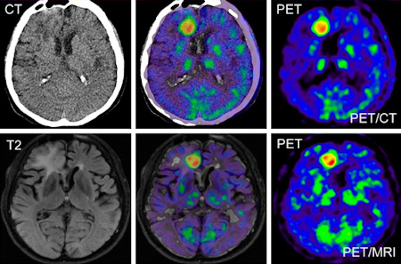
Figure 9: CT, T2, PET and CT/PET, T2/PET of a GBM [25]
Further reading
A recent study by researchers from the University of Michigan Medical School and Harvard University shows good results for tumor diagnosis via a combination of SRS (Stimulated Raman Scattering) microscopy and deep learning. The paper related to the study can be found here.
Bibliography
[2] http://emedicine.medscape.com/article/1147993-overview#a2
[3] http://www.abta.org/brain-tumor-information/diagnosis/biopsy-procedure.html
[4] http://www.abta.org/brain-tumor-information/diagnosis/laboratory-tests.html
[5] http://www.abta.org/brain-tumor-information/diagnosis/dna-profiling.html
[6] http://abc2.org/guidance/10-steps/10-test-your-tumors-genetic-makeup
[8] http://www.abta.org/brain-tumor-information/diagnosis/types-of-brain-scans.html
[11] http://web.mit.edu/kitmitmeg/whatis.html
[13] https://www.ndcn.ox.ac.uk/divisions/fmrib/what-is-fmri/introduction-to-fmri
[14] http://www.mayfieldclinic.com/PE-SPECT.htm
[15] http://www.mayfieldclinic.com/PE-MRspectroscopy.htm
[19] http://www.keywordsuggests.com/Qe26decSKaUW6zHHe98lyOUrsVSUlYQ0L6*%7C59NgeLc/
[21] http://emedicine.medscape.com/article/2110836-overview
[22] http://casemed.case.edu/clerkships/neurology/Web%20Neurorad/MRI%20Basics.htm
[23] https://en.wikipedia.org/wiki/Magnetic_resonance_angiography
[25] http://www.auntminnie.com/index.aspx?sec=ser&sub=def&pag=dis&ItemID=91503
2 Kommentare
Unbekannter Benutzer (gu95wiq) sagt:
16. Mai 2017Very good and short overview over all diagnosis modalities.
Especially, I like the further readings addition, thank you!
One thing: A Bibliography with all the links would be advantageous!
Otherwise everything is fine, nice work!
Unbekannter Benutzer (ga67yur) sagt:
16. Mai 2017Thanks for the feedback! And yes, we will work on the bibliography.