Daniel Berger, winter semester 2015/16
The method’s purpose
X-rays are electromagnetic waves with very short wavelengths, which spread out with speed of light and can penetrate basically every material. Radiations passing through a test object can be detected and thereof two-dimensional projections of the inner structure of an object can be generated. Computed tomography (CT) extends the technique by generating three-dimensional virtual reconstructions and sectional images out of a set of two-dimensional projections from different angular perspectives.
Historical Background
X-rays have been discovered unintentionally by W.C. Roentgen in 1895 by experimental tests with a gas discharge tube as black photographic paper emitted visible light because of fluorescence. Thereafter, A.H. Becquerel, Marie Curie and Pierre Curie discovered the radioactivity of uranium ore, polonium and radium. Based on these findings, X-rays have been researched step by step by many well-known scientists. X-ray diffraction on crystals, characteristic X-ray spectrums of different elements, interactions of X-rays and electrons, diffractions of X-rays and electrons in gases and CT (1979) have been milestones of the research’s development. Moreover, the continuous progress of X-rays enabled microstructural researches on e.g. DNA, proteins and crystals. Several Nobel Prize awards emphasized the enormous achievements in radiography during the last century.[1][2][3]
Physical basics of X- and gamma rays
Wave radiation
X- and gamma rays are electromagnetic waves, which have, according to the quantum theory, neither masses nor electric charges. First of all, they are characterized by their radiant energy. The wavelength λ and the frequency f are the most important quantities, which correlate with each other via the propagation speed (speed of light c) as follows:
{c=λ f}.
The radiant energy E is defined by the Planck constant h and the frequency f:
E=hf.
The typical radiant energy is in the range of E = [10 keV; 10 MeV] and the wavelength in the range of λ = [10^{-8} m; 10^{-13} m]. The shorter the wavelengths the harder and stronger are the associated rays.
For historical reasons the term “gamma ray” is often used for natural -sources. In this case, the radiant energy depends on the type and the size of the source.[4]
X-rays
Usually, highly accelerated electrons striking on materials generate X-rays. Provided that the energy is ideally transferred, the resulting photon energy is equal to the kinetic energy of the incident electron.
E_{kin}=E_{max}=\frac{1}{2} m v^2=h f_{max}(=e U_a=h\frac{c}{λ_{min}}).
This illustrates the significant influence of the accelerated electrons on the resulting rays. There are two basic modes of X-ray generation, namely the braking and the characteristic radiation.[3]
Braking radiation
Braking radiation is generated by accelerated electrons (10 to 99% of speed of light), which enter the atomic shell of certain materials. The electrons loose their kinetic energy and thereby photon quants are emitted. Depending on how fast the electrons are decelerated, X-rays with different wavelengths in a continuous spectrum are generated. At a certain minimal wavelength λ_{min}, an electron transfers its kinetic energy completely to the generated photon quant. Higher velocities of the striking electrons solely are able to reduce this critical wavelength. Furthermore, this value is a decisive quantity for the intensity and penetration ability of the radiation. It is calculated as follows:
{λ_{min}=\frac{h c}{e U_A}}.
h: Planck constant (6,626070040 \cdot 10^{-34}J s)
e: elementary electric charge (1,6021766208 \cdot10^{-19}C)
c: speed of light (299792458\frac{m}{s})
U_A: acceleration voltage[3]
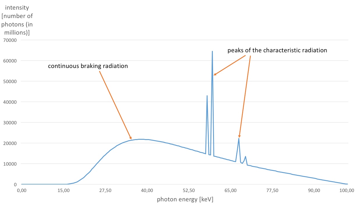
A typical example of an element spectrum is given in figure 1.
| Figure 1: Simulated element spectrum of a tungsten anode (acceleration voltage 100 kV). |
Characteristic radiation
This radiation is comparatively much more intensive than the braking radiation. Striking electrons as well generate it. In this case the entering electrons separate electrons of the atomic shell and ionize atoms. Consequently, an electron of another energy level (typically the next higher one) skips to the free spot and a defined amount of energy is emitted in the form of photon quants. These shell-transitions can continue in cascades. Depending on the stroked chemical elements they produce emission spectrums with characteristic peaks at certain wavelengths (see figure 1). Characteristic radiation is also called K-shell emission as the emission at the atomic K-shell always represents the resulting radiation with the highest energy level.[1][2][3]
Properties of X-rays
- Located outside of the visible spectrum
- No possibility of influencing by electric of magnetic fields
- Possibility of ionization by separating electrons from the atomic shell
- Linear spreading with speed of light
- Diffraction at boundary layers of different materials
- Attenuated after striking on materials
- emission of visible light caused by fluorescence
- Chemical and biological effects on (an-)organic materials – particularly potential damage of somatic cells
- Ability of penetrating basically every kind of material[5][2]
Verification of the presence of X-rays
This electromagnetic radiation can be detected differently. Two of the most common detection principles are described now:
Ionization chambers are filled with gas and have two electrodes, which are applied with an acceleration voltage. Due to the radiation, the gas is partly ionized and the gas disintegrates into ions and electrons. Thereafter, they are accelerated to the corresponding electrode and generate a test pulse caused by the strike. The frequency of the test pulses correlates to the energy dose of the radiation. It needs to be paid attention that the acceleration voltage is high enough in order to eliminate reintegration of ions and electrons. This voltage is called saturation voltage.
In principle the assembly of the Geiger-Mueller counter is equal to the ionization chamber. However, the acceleration voltage is much higher, which strengthens the test pulses and makes it possible to count the charged particles. Many devices instantly indicate these pulses by a cracking noise. Furthermore, it’s possible to determine the range, the penetrating ability and the type of the radiation by positioning an object between the radiation source and the counter.[6]
Dosimetric quantities
The energy dose of X-rays is defined as follows:
energy\ dose\ D = \frac{absorbed\ energy\ E}{tissue\ mass\ M}.
For this physical parameter, the following units can be used:
1\ Gy = 1 \frac{m^2}{s^2} = 1 \frac{J}{kg} = 10^2 rad.
Furthermore, the energy dose rate is defined by:
energy\ dose\ rate\ R = \frac{absorbed\ energy\ E}{tissue\ mass\ M \cdot time\ t} (1\frac{rad}{s}= 10^{-2} \frac{Watt}{kg}).
Information with regard to the radiation intensity can alternatively be provided by the ion dose. However, it is defined for the special case of air-ionization only. This also applies to the ion dose rate, which gives information about the ion dose being absorbed in a certain time interval.
The equivalent dose and the effective equivalent dose (SI-unit Sievert/Sv) are further relevant dosimetric quantities.[7][6]
Implementation of the X-ray technologiy in non-destructive testing
The described physical basics in the upper sections are mainly applied in medicine (diagnostic, nuclear therapy) and in component testing. In this section, the most important steps and factors for the generation of X-ray images are described. In addition, information about different X-ray tubes and devices and the possibilities of computed tomography will be provided.
Generation of X-ray images
The generation of X-ray images requires a certain system structure. Moreover, the applier needs to have basic knowledge about the physical interaction mechanisms of X-rays with matter and the detection of X-rays, both needed to correctly interpret the results.
Structure of X-ray devices
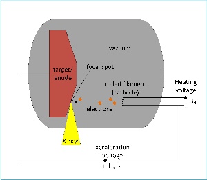
The artificial generation of X-rays usually takes place in vacuum tubes (see figure 2). A coiled filament at the cathode (in most cases tungsten) being electrically heated up to around 2400 K releases electrons through thermionic emission. The electrons are accelerated to the anode as a voltage potential Ua between the cathode and the anode is applied. Depending on the required X-ray intensity, this voltage is typically in the range of 25 kV to 500 kV. The electrons strike on the anode disk and generate the X-rays. As approximately 99% of the kinetic energy of the electrons will be converted into heat, the anode material requires very high values for melting point and thermal conductivity. Molybdenum alloys and tungsten composites fulfill these needs. In addition to that, the anode disk can be turned continuously during the radiation process to distribute the generated heat to a larger surface area.
Basically there are two different types of X-ray tubes:
- Reflection tube: The accelerated electrons strike on a chamfered target (anode) and generate the X-rays, which leave the anode disk conical in another angle than the original electron beam.
- Transmission tube: The electrons strike orthogonal on a very thin anode plate (e.g. tungsten). The generated X-rays penetrate the target and leave the tube aligning to the electron beam.
As gamma devices use naturally emitting gamma sources it is not necessary to explain the gamma-ray generation. In this case the focusing of the gamma-ray beam and the radiation protection with shielding is more important. The gamma source can be driven in a save position or working position via remote control, which excludes unintentional irradiation of the environment or the applier himself. This sort of application typically uses disintegrating isotopes like e.g. iridium 192, cobalt 60 or tantalum 182.[4]
| Figure 2: Schematic representation of an X-ray tube. |
Penetrating ability
The energy dose of X-rays is reduced when passing through matter. This process mainly depends on the chemical and physical properties of the irradiated material and the original energy dose of the X-rays. The Lambert-Beer’s law describes this reduction:
P(x)=P_0e^{-μx}.
P_0: original lintensity
x: material thickness
μ: attenuation coefficient
The half-value layer is a physical variable that provides information about the penetration ability. It is the thickness of a material, which is necessary to halve the intensity of X-rays with a certain energy dose.
The reduction of the radiation intensity takes place at the atomic level and is based on different mechanisms of attenuation, which are described below. The mechanisms are one reason for blurring in the resulting image, as they produce scattered X-ray photons.
Photoelectric effect: An electron of the atomic shell absorbs the entire energy of a striking photon. Due to this energy the electron is released and leaves the atomic shell and an electron from a higher energy level skips to the free spot. During this process, called X-ray fluorescence, characteristic radiation is generated and transmitted.
Compton scattering: This effect describes the interaction between an electron of the atomic shell and a striking photon. In this case the photon isn’t deleted but scattered and spreads in some other direction with a longer wavelength as it looses energy during the interaction. Depending on the amount of the energy being transferred to the electron, it stays on its position in the atomic shell or is emitted equal to the photoelectric effect.
Elastic Rayleigh scattering: The photon is scattered on the atomic shell. In this process no energy is transferred and following the photon moves in another direction with the same wavelength as before.
Pair production: The photon disintegrates into an electron and a positron, which move in different and new directions. The original kinetic energy is divided across the particles. The electron is now able to initiate a second ionization. As the effect can only be caused by photons with an energy that is higher than 1,02 MeV, it exclusively appears when using gamma emitters.
Apart from these major effects there are several other nuclear reactions, which lead to emissions of neutrons and protons.[4][2][6]
Radiographic detection
Radiographic detection is generally divided into the conventional analogue film radiography and the digital radiography:
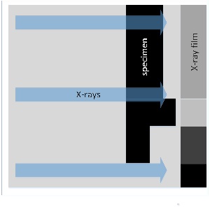
Analogue film radiography: Small AgBr-grains on the surface of the film react to X-rays with disintegration. After the radiographic film development, they appear darker in visible light. This generates a contrast between parts where X-rays with higher intensity stroked on the film and others. The most important disadvantage of this technique is the light sensitivity and the development of the film. The principle is shown in figure 3.[2][8][6]
Digital radiography: The digital detector technology uses different methods to create radiographic images. For example, phosphorous screens, which react to X-rays with emission of visible light that is recorded with a video camera, are used. There are some more methods, which all are similar to the Geiger-Mueller counter. Indirect test pulses, which are based on ionization, can be recorded and counted on a two-dimensional array of micro electric sensors. This type of detectors is mainly characterized by the following factors:
- Spatial resolution/detection efficiency: How much energy does a photon need to have to provoke a measurable test pulse?
- Linearity: Is the amount of test pulses measured on the detector proportional to the amount of the photon quants? The dead time is very important for this factor as it describes the time between two test pulses, in which the detector isn’t able to recognize any pulse.
- Proportionality: Is there a linear relation between the photon energy and the test pulses?
- Dynamic range: The interval of intensity between minimum detection and saturation.
- Contrast: The ability of the system to distinguish close structures with similar attenuation properties (for example fat-muscle)[4][2][3]
In addition, there are other types of digital detectors, as for example, imaging plates, charge-coupled-devices (CCD) and flat panel detectors (FPD).
The digitalization significantly reduced the effort of generating and administrating X-ray images. Typical representatives of this technology are CT, computed radiography (CR), real-time radiography and direct radiography.[8]
| Figure 3: Schematic representation of an X-ray system with X-rays, specimen and X-ray film. |
Imaging quality
The main factors, which influence imaging quality, are contrast and blurring:
The contrast is the difference of grayscale between an object and its environment on an X-ray image. It is influenced by the properties of the X-rays, the energy dose, the quality of the detector the properties of the specimen.
The geometric blurring stands for the influence of geometric effects in the system assembly on the imaging quality. The most important correlations are as follows:
- the smaller the distance between object and detector as well as the longer the distance between the object and the focal spot, the sharper and smaller is the image
- the smaller the focal spot, the is sharper the image
The internal blurring is the impact with regard to the imaging quality of the AgBr-grain size on the surface of analogue radiographic films. The principle is, the smaller the grains, the sharper the image. However, very small grains necessarily require the extension of the radiation exposure time, as they disintegrate slower than large grains.
This is of considerable relevance, particularly in medical applications, where there is always the objective to reduce the dose for the patient.
Further impact factors on the imaging quality are the movement of test object, film and X-ray tube during the process and the combination of film and film coating in analogue radiography.
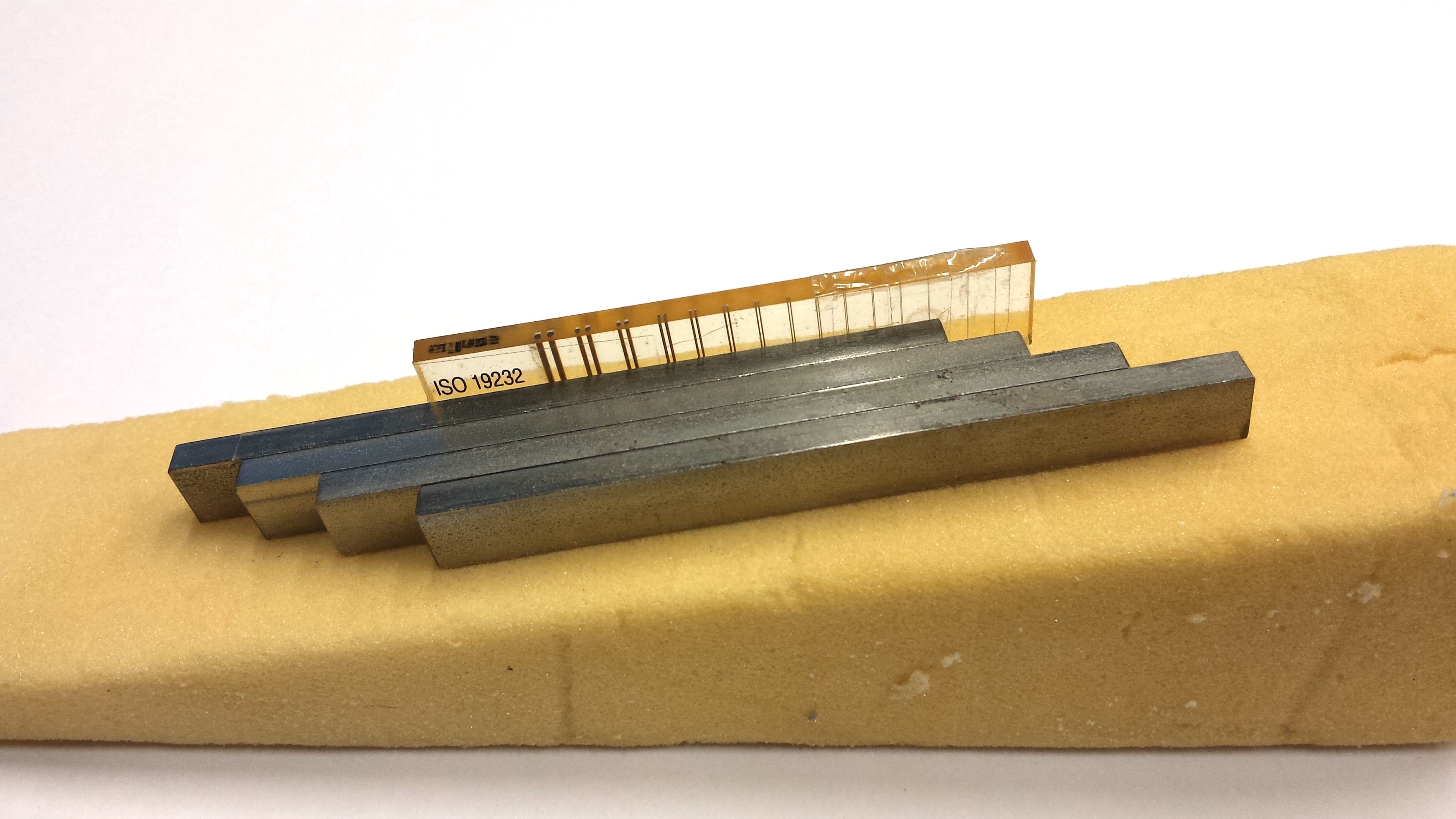
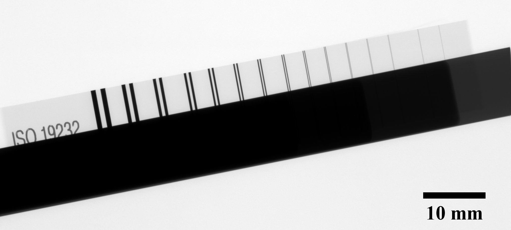
Imaging quality can be measured with special test specimen, so-called wire penetrameter plates. These are thin wires with different predefined thicknesses, which are integrated into small plastic plates. They are radiographically recorded together with the test object. This finally allows finding out to which minimal degree of thickness an object generating contrast in the image can be reliably recognized. In figure 4 and 5 an example of the plate and an X-Ray projection are shown.[5][4][2][8][6]
| Figure 4: Example of a wire penetramter plate (duplex wire type Q) with test objects. | Figure 5: Example of an X-ray projection of test objects in figure 4. |
Computed tomography
Computed tomography (tomos: slice and graphein: draw) is a high-tech digital sectional imaging method having its roots from the 20’s.
The idea of the method is to create several X-ray images from different perspectives. The resulting data is merged together with complex software tools into volume elements (so-called voxels). This allows generating three-dimensional surface reconstructions being displayed in a virtual space. The technique is mainly used for presentation purposes. As the filtering during the process can cause loss of information, experts usually use two-dimensional sectional images for evaluation, which can be deduced from the voxel data as well. The major advantage of these images is that they don’t show averaged values being generated automatically by a considerable reduction of the beam intensity in conventional two-dimensional X-ray images. Summed up, computed tomography allows generating detailed morphologic virtual reproductions of parts of a body or other specimen. Furthermore, size-measurements on material faults can be carried out with this method.
In medicine CT-devices are usually designed to allow rotational movements of the X-ray tube and the detector around the test object. The fastest devices turn round once in 0.33 seconds. This enormous velocity combined with other techniques like multi-detector systems, simultaneous use of several X-ray tubes or special devices for heartbeat-synchronization in cardiologic examinations, exclude nearly every artefact based on movements of the living test object.[4][3][9][6]
Radiation protection
X-rays particularly have injurious effects on biologic cellular tissue and DNA chains. In the worst cases genetic information can be damaged irreversibly or stem cells can be destroyed. Although, every living being has a natural protection system against nuclear radiation, these biological mechanisms are not able to compensate the energy doses of technical radiation. Due to the intensity of these energy doses, X-rays in medical devices are restricted by law to protect appliers and patients.
I.a., radiation protection includes the shielding of radiators to avoid unintentional irradiation of the environment and the applier. Materials with very short half-value layers are used to focus the beam of radiation or to obscure the radiant completely (in particular gamma-sources). To minimize the radiation exposure, remote controls and additional lead-shields for very sensitive and irrelevant areas of the body are used.
Moreover, there are methods like intensifying screens that reduce the energy dose needed to generate meaningful X-ray images. During the analogue film radiography, they are positioned in front of and behind of the film. The screens react on X-rays with light emission and by this strengthen the film density. Consequently, the energy dose can be reduced by around 95%.
The Newton’s inverse square law is a phenomenon of radiation protection occurring naturally. Caused by the divergence of the radiation its intensity decreases with higher distances to the radiator as follows:
I_1=I_0 \cdot \frac{r_0}{r_1^2}.
r_0: distance 0 to focal spot of radiant:
r_1: distance 1 to focal spot of radiant
I_0: radiation intensity at r_0
I_1: radiation intensity at distance r_1
This means, that if the distance to the focal spot of the radiant is doubled, the energy dose per area is reduced to \frac{1}{4}.[1][6]
Application examples in material testing and medicine
In the field of non-destructive testing, radiographic examinations are primarily used to indicate and locate macroscopic volumetric defects like foreign inclusions, pores, segregations, different thicknesses and filling material. Typically, safety-relevant cast parts or parts with welding joints are examined. Additionally, CT methods enable the size-measurement of defects, which can be used to compute the durability of the examined part. Another benefit of the CT is the detection of inner cracks as the identification is hardly possible with conventional two-dimensional X-ray methods in case that the crack direction is similar to the X-rays.
The microradiography is an additional field of application where thin metal plates are penetrated with very hard X-rays. The results of the examination allow conclusions about the size and the form of the material’s microstructure.
Medical radiologic examinations principally serve for more objective clinic diagnoses, scientific researches or the support of expert’s reports. Predominantly X-rays help to assess malfunctions of the musculoskeletal system. This includes structural analyses to detect or to exclude causes of malfunctions and to observe changes during courses of disease. Typical examinations of the human body are the functional analysis of the spinal column, situation and error analyses of the jaw area, joint problems and bone fractures. Furthermore, medical X-ray images are used to train prospective doctors. With the help of the technology basic knowledge about the musculoskeletal system is mediated.[5][9]
Literature
- Eichler, Hans J., Kronfeldt, Heinz-Detler, Sahm Jürgen: Das Neue Physikalische Grundpraktikum. Springer-Verlag Berlin Heidelberg (2001), p. 477-487
- Große, Christian U.: Einführung in die Zerstörungsfreie Prüfung im Ingenieurwesen. Grundlagen und Anwendungsbeispiele. Version 2015-07-01, Literatur:
Aichinger, H. et al.: Radiation exposure and image quality in x-ray diagnostic radiologiy, Springer Verlag, Berlin, 2012, ISBN: 3642112412
Askeland, D.R.: Materialwissenschaften, Spektrum Akademischer Verlag, Heidelberg, 1996
Cartz, L.: Nondestructive Tesing – Radiography, Ultrasonics, Liquid Penetrant, Magnetic Particle, Eddy Current, 1995, ASM Intl; ISBN: 0871705176
Erhardt, A.: Verfahren der Zerstörungsfreien Materialprüfung – Grundlagen. 2014; Hrsg. DGZfP; DVS Media GmbH.
Heine, B.: Werkstoffprüfung – Ermittlung von Werkstoffeigenschaften, Fachbuchverlag Leipzig, 2003, ISBN: 3446-222847
Kak, A.C. und Slaney, M.: Principles of computerized tomographic imaging, New York: IEEE Press, 1987
Quinn, R.: Radiography in modern industry. Fourth Edition, Eastman Kodak Company, Rochester, New York, 1980
Puschke, M.: Die Röntgenprüfung, Castell-Verlag GmbH, Wuppertal, 2001, ISBN: 3934225071
Schönen, J.: Ionisierende Strahlung, Strahlungsarten und ihre Wechselwirkungen mit Materie, 2001
Wuich, W.: Zerstörungsfreie Werkstoffprüfung, Band 5, Verlag Erwin Greyer, Bad Wörishofen, 1971
Introduction To Radiation: The Health Physics Society, The University of Michigan - Spieß, Lothar, Teichert, Gerd, Schwarzer, Robert, Behnken, Herfried, Christoph Genzel: Moderne Röntgenbeugung. Röntgendiffraktometrie für Materialwissenschaftler, Physiker und Chemiker. 2. Auflage Vieweg+Teubner | GWV Fachverlage GmbH Fachverlage GmbH, Wiesbaden (2009), p. 5-39, p. 95-153
- Buzug, Thorsten M. Computed Tomography. From Photon Statistics to Modern Cone-Beam CT. Springer-Verlag Berlin Heidelberg (2008), p. 1-52, p. 75-98
- Berthold, R., Vaupel O., Förster F., Siebel E. (Hrsg.), Ludwig N. (Hrsg.): Handbuch der Werkstoffprüfung. Erster Band. Prüf- und Messeinrichtungen. Springer-Verlag Berlin Heidelberg GmbH (1985), p. 576-608
- Vogl, Thomas J., Reith, Wolfgang, Rummeny, Ernst J. (Hrsg.): Diagnostische und Interventionelle Radiologie. Springer-Verlag Berlin Heidelberg (2011), p. 4-35
- Pollermann Max, Gerthsen: Einführung in das Pysikalische Praktikum. Für Mediziner und für das Anfängerpraktikum. Springer-Verlag, Berlin Heidelberg (1967), p. 145-171, p. 1-52, p. 75-98
- Pacella Danilo, Energy-resolved X-ray detectors: the future of diagnostic imaging. ENEA-Frascati, Rome, Eval. (2014), p. 1-14
- Tilscher, H., Graf E.: Die Bedeutung der bildgebenden Verfahren – Röntgen, CT und MRT – in der konservativen Orthopädie und manuellen Medizin. Springer-Verlag (2010), p. 16-22

